Sunday Poster Session
Category: Liver
P1416 - Unveiling Silent Infiltration: A Case Study of Jaundice and Breast Cancer
Sunday, October 27, 2024
3:30 PM - 7:00 PM ET
Location: Exhibit Hall E

Has Audio

Lakmal Ekanayake, DO
Wright State University Boonshoft School of Medicine
Centerville, OH
Presenting Author(s)
Lakmal Ekanayake, DO1, Adam Myer, MD2, Benjamin Clement, MD2, Kelsey Muchmore, PA-C2, Askanda Osman, MD2, Amoah Yeboah-Korang, MD3
1Wright State University Boonshoft School of Medicine, Centerville, OH; 2University of Cincinnati Medical Center, Cincinnati, OH; 3University of Cincinnati Medical Center, CIncinnati, OH
Introduction: Metastatic disease to the liver is a known site of breast cancer metastases, typically presenting as hypovascular or hypervascular masses within the liver1. In very rare cases, hepatic metastatic disease presents as intra-sinusoidal infiltration undetectable radiographically.1
Case Description/Methods: A 66-year-old Caucasian female presented with jaundice, unintentional weight loss, and left facial droop. Past medical history included stage 3 invasive lobular breast cancer 4 years ago, estrogen receptor positive, progesterone receptor positive, and HER2 negative status post mastectomy with negative margins but 14 of 14 positive lymph nodes. Patient was maintained on aromatase inhibitors with sustained clinical and radiographic remission.
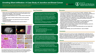
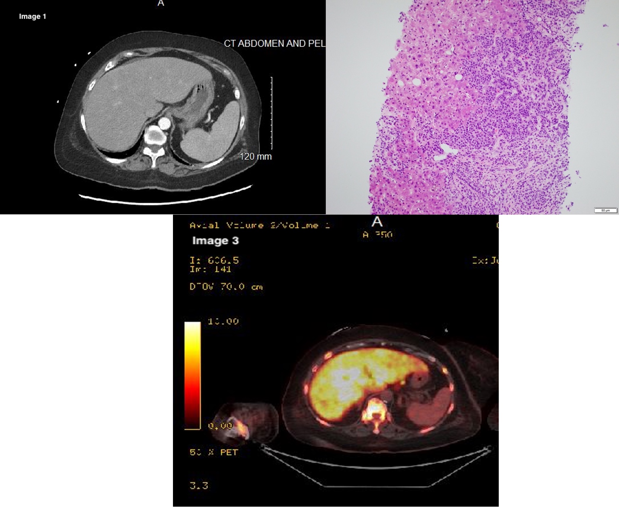
Physical exam showed jaundice and left facial droop. Liver tests were notable for elevated total bilirubin 16.6 (direct 8.83), alkaline phosphatase 565, AST 176 and ALT 80. CT abdomen, pelvis with contrast was unremarkable [Image 1]. MRCP showed scattered areas of peripheral biliary ductal dilatation and central attenuation consistent with cholangitis; no evidence of metastatic disease in the liver. Viral and autoimmune serologies were unremarkable. MRI brain was unrevealing for etiology of facial droop. Liver biopsy showed poorly differentiated carcinoma, consistent with metastasis from known breast primary [Image 2], CK7 (positive), GATA-3 (positive), and GCDFP focal positive. Whole-body PET CT revealed widespread axial and appendicular skeletal metastases in addition to diffuse liver uptake which is consistent with known metastatic disease [ Image 3].
Discussion: Metastatic breast cancer to the liver not seen radiographically is extremely rare; however, it should be suspected in patients with a history of breast cancer and unexplained jaundice. Liver biopsy and PET CT are required for diagnosis and staging.

Disclosures:
Lakmal Ekanayake, DO1, Adam Myer, MD2, Benjamin Clement, MD2, Kelsey Muchmore, PA-C2, Askanda Osman, MD2, Amoah Yeboah-Korang, MD3. P1416 - Unveiling Silent Infiltration: A Case Study of Jaundice and Breast Cancer, ACG 2024 Annual Scientific Meeting Abstracts. Philadelphia, PA: American College of Gastroenterology.
1Wright State University Boonshoft School of Medicine, Centerville, OH; 2University of Cincinnati Medical Center, Cincinnati, OH; 3University of Cincinnati Medical Center, CIncinnati, OH
Introduction: Metastatic disease to the liver is a known site of breast cancer metastases, typically presenting as hypovascular or hypervascular masses within the liver1. In very rare cases, hepatic metastatic disease presents as intra-sinusoidal infiltration undetectable radiographically.1
Case Description/Methods: A 66-year-old Caucasian female presented with jaundice, unintentional weight loss, and left facial droop. Past medical history included stage 3 invasive lobular breast cancer 4 years ago, estrogen receptor positive, progesterone receptor positive, and HER2 negative status post mastectomy with negative margins but 14 of 14 positive lymph nodes. Patient was maintained on aromatase inhibitors with sustained clinical and radiographic remission.
Physical exam showed jaundice and left facial droop. Liver tests were notable for elevated total bilirubin 16.6 (direct 8.83), alkaline phosphatase 565, AST 176 and ALT 80. CT abdomen, pelvis with contrast was unremarkable [Image 1]. MRCP showed scattered areas of peripheral biliary ductal dilatation and central attenuation consistent with cholangitis; no evidence of metastatic disease in the liver. Viral and autoimmune serologies were unremarkable. MRI brain was unrevealing for etiology of facial droop. Liver biopsy showed poorly differentiated carcinoma, consistent with metastasis from known breast primary [Image 2], CK7 (positive), GATA-3 (positive), and GCDFP focal positive. Whole-body PET CT revealed widespread axial and appendicular skeletal metastases in addition to diffuse liver uptake which is consistent with known metastatic disease [ Image 3].
Discussion: Metastatic breast cancer to the liver not seen radiographically is extremely rare; however, it should be suspected in patients with a history of breast cancer and unexplained jaundice. Liver biopsy and PET CT are required for diagnosis and staging.

Figure: Image 1 (Left, Top)1: CT Abdomen and Pelvis. Image 2 ( Right, Top): Liver Biopsy. Image 3 (Bottom): PET-CT scan, Liver uptake
Disclosures:
Lakmal Ekanayake indicated no relevant financial relationships.
Adam Myer indicated no relevant financial relationships.
Benjamin Clement indicated no relevant financial relationships.
Kelsey Muchmore indicated no relevant financial relationships.
Askanda Osman indicated no relevant financial relationships.
Amoah Yeboah-Korang indicated no relevant financial relationships.
Lakmal Ekanayake, DO1, Adam Myer, MD2, Benjamin Clement, MD2, Kelsey Muchmore, PA-C2, Askanda Osman, MD2, Amoah Yeboah-Korang, MD3. P1416 - Unveiling Silent Infiltration: A Case Study of Jaundice and Breast Cancer, ACG 2024 Annual Scientific Meeting Abstracts. Philadelphia, PA: American College of Gastroenterology.
