Sunday Poster Session
Category: Pediatrics
P1477 - Hepatomegaly and Recurrent Cholangitis in an Adolescent Male
Sunday, October 27, 2024
3:30 PM - 7:00 PM ET
Location: Exhibit Hall E

Has Audio

Nguyen Pham, MD
Ronald Reagan Hospital
Los Angeles, CA
Presenting Author(s)
Nguyen Pham, MD1, Keiko Yoshioka, DO2, Sarah Gustafson, MD3
1Ronald Reagan Hospital, Los Angeles, CA; 2Harbor-UCLA Medical Center, Torrance, CA; 3Harbor-UCLA Pediatrics, UCLA David Geffen School of Medicine, Torrance, CA
Introduction: Hepatomegaly is common in the pediatric population and the etiologies of hepatomegaly can be categorized into six large domains such as infectious, inflammatory, autoimmune, congenital, neoplastic disorders and structural. Caroli’s disease is a rare, frequently missed structural etiology of hepatomegaly. We present a rare case of Caroli’s disease in an adolescent male with recurrent cholangitis and hepatomegaly.
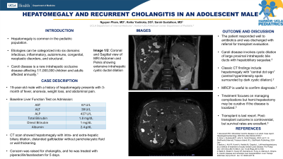
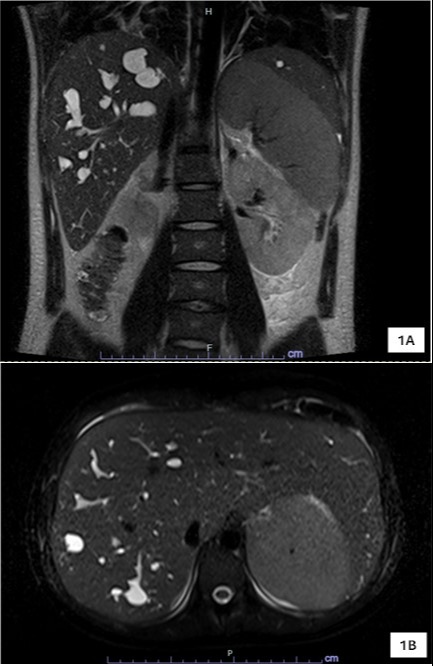
Case Description/Methods: A 19-year-old male with a history of hepatomegaly presents with a 3-month of fevers, night sweats, anorexia, melena, emesis, weight loss and upper abdominal pain. His vitals were normal and exam was remarkable for left upper quadrant abdominal tenderness, hepatomegaly and splenomegaly. Laboratory results showed elevated alkaline phosphatase, transaminase and conjugated hyperbilirubinemia. CT abdomen/pelvis with contrast showed hepatomegaly with intra- and extra-hepatic biliary dilation, distended gallbladder without pericholecystic fluid or wall thickening, mild splenomegaly. A magnetic resonance cholangiopancreatography (MRCP) showed irregular intra- and extra-hepatic stricture of the biliary system including common hepatic duct with fusiform and peripheral saccular dilations of the liver (Image 1a and 1b) consistent with Caroli’s disease. He was treated for cholangitis with a 5-day course of piperacillin/tazobactam and discharged with referral for liver transplant evaluation at a tertiary center.
Discussion: Caroli disease is a rare intrahepatic occlusive disease associated with autosomal recessive polycystic kidney disease. The disease involves cystic dilation of large proximal intrahepatic bile ducts complicated by cholangitis, hepatic dysfunction and non-cirrhotic portal hypertension. Classic CT findings includes hepatomegaly with “central dot sign” representing central hyperintensity spots surrounded by dark cystic dilation along with intrahepatic and/or extrahepatic biliary dilation . MRCP may show non-obstructive cystic biliary dilation. Treatment of Caroli’s disease focuses on managing its complications but hemi-hepatectomy may be curative if the disease is localized. Advanced cases may require liver transplantation but post-transplantation outcome is controversial. This case highlights the importance of early recognition of Caroli’s disease in patients with hepatomegaly of unknown etiology and recurrent cholangitis. Early recognition is important for prevention of complications and appropriate specialty care for liver transplant evaluation.

Disclosures:
Nguyen Pham, MD1, Keiko Yoshioka, DO2, Sarah Gustafson, MD3. P1477 - Hepatomegaly and Recurrent Cholangitis in an Adolescent Male, ACG 2024 Annual Scientific Meeting Abstracts. Philadelphia, PA: American College of Gastroenterology.
1Ronald Reagan Hospital, Los Angeles, CA; 2Harbor-UCLA Medical Center, Torrance, CA; 3Harbor-UCLA Pediatrics, UCLA David Geffen School of Medicine, Torrance, CA
Introduction: Hepatomegaly is common in the pediatric population and the etiologies of hepatomegaly can be categorized into six large domains such as infectious, inflammatory, autoimmune, congenital, neoplastic disorders and structural. Caroli’s disease is a rare, frequently missed structural etiology of hepatomegaly. We present a rare case of Caroli’s disease in an adolescent male with recurrent cholangitis and hepatomegaly.
Case Description/Methods: A 19-year-old male with a history of hepatomegaly presents with a 3-month of fevers, night sweats, anorexia, melena, emesis, weight loss and upper abdominal pain. His vitals were normal and exam was remarkable for left upper quadrant abdominal tenderness, hepatomegaly and splenomegaly. Laboratory results showed elevated alkaline phosphatase, transaminase and conjugated hyperbilirubinemia. CT abdomen/pelvis with contrast showed hepatomegaly with intra- and extra-hepatic biliary dilation, distended gallbladder without pericholecystic fluid or wall thickening, mild splenomegaly. A magnetic resonance cholangiopancreatography (MRCP) showed irregular intra- and extra-hepatic stricture of the biliary system including common hepatic duct with fusiform and peripheral saccular dilations of the liver (Image 1a and 1b) consistent with Caroli’s disease. He was treated for cholangitis with a 5-day course of piperacillin/tazobactam and discharged with referral for liver transplant evaluation at a tertiary center.
Discussion: Caroli disease is a rare intrahepatic occlusive disease associated with autosomal recessive polycystic kidney disease. The disease involves cystic dilation of large proximal intrahepatic bile ducts complicated by cholangitis, hepatic dysfunction and non-cirrhotic portal hypertension. Classic CT findings includes hepatomegaly with “central dot sign” representing central hyperintensity spots surrounded by dark cystic dilation along with intrahepatic and/or extrahepatic biliary dilation . MRCP may show non-obstructive cystic biliary dilation. Treatment of Caroli’s disease focuses on managing its complications but hemi-hepatectomy may be curative if the disease is localized. Advanced cases may require liver transplantation but post-transplantation outcome is controversial. This case highlights the importance of early recognition of Caroli’s disease in patients with hepatomegaly of unknown etiology and recurrent cholangitis. Early recognition is important for prevention of complications and appropriate specialty care for liver transplant evaluation.

Figure: Image 1A - Coronal View of MRI Abdomen and Pelvis showing extensive intrahepatic cystic ductal dilation; Image 1B - Sagittal View of MRI Abdomen and Pelvis showing a different perspective of the intrahepatic cystic ductal dilation.
Disclosures:
Nguyen Pham indicated no relevant financial relationships.
Keiko Yoshioka indicated no relevant financial relationships.
Sarah Gustafson indicated no relevant financial relationships.
Nguyen Pham, MD1, Keiko Yoshioka, DO2, Sarah Gustafson, MD3. P1477 - Hepatomegaly and Recurrent Cholangitis in an Adolescent Male, ACG 2024 Annual Scientific Meeting Abstracts. Philadelphia, PA: American College of Gastroenterology.
