Sunday Poster Session
Category: Pediatrics
P1478 - Enteric Duplication Cysts: An Overlooked Cause of Recurrent Pancreatitis
Sunday, October 27, 2024
3:30 PM - 7:00 PM ET
Location: Exhibit Hall E

Has Audio

Palack Agrawal, MD
Memorial Healthcare System
Hollywood, FL
Presenting Author(s)
Palack Agrawal, MD, Keshav Bhattar, MD, Claudia Rojas, MD, Diego Diaz, MD
Memorial Healthcare System, Hollywood, FL
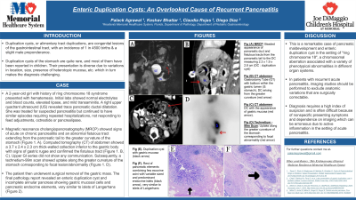
Introduction: Duplication cysts, or alimentary tract duplications, are congenital lesions of the gastrointestinal tract, with an incidence of 1 in 4500 births & a slight male preponderance. Duplication cysts of the stomach are quite rare, and most of them have been reported in children. Their presentation is diverse due to variations in location, size, presence of heterotopic mucosa, etc. which in turn makes the diagnosis challenging.
Case Description/Methods: A 2-year-old girl with history of ring chromosome 18 syndrome presented with hematemesis. Initial labs showed normal electrolytes and blood counts, elevated lipase, and mild transaminitis. A right upper quadrant ultrasound (US) revealed trace pancreatic ductal dilatation. She was treated for suspected pancreatitis but continued to have similar episodes requiring repeated hospitalisations, not responding to feed adjustments, octreotide or pancrealipase.
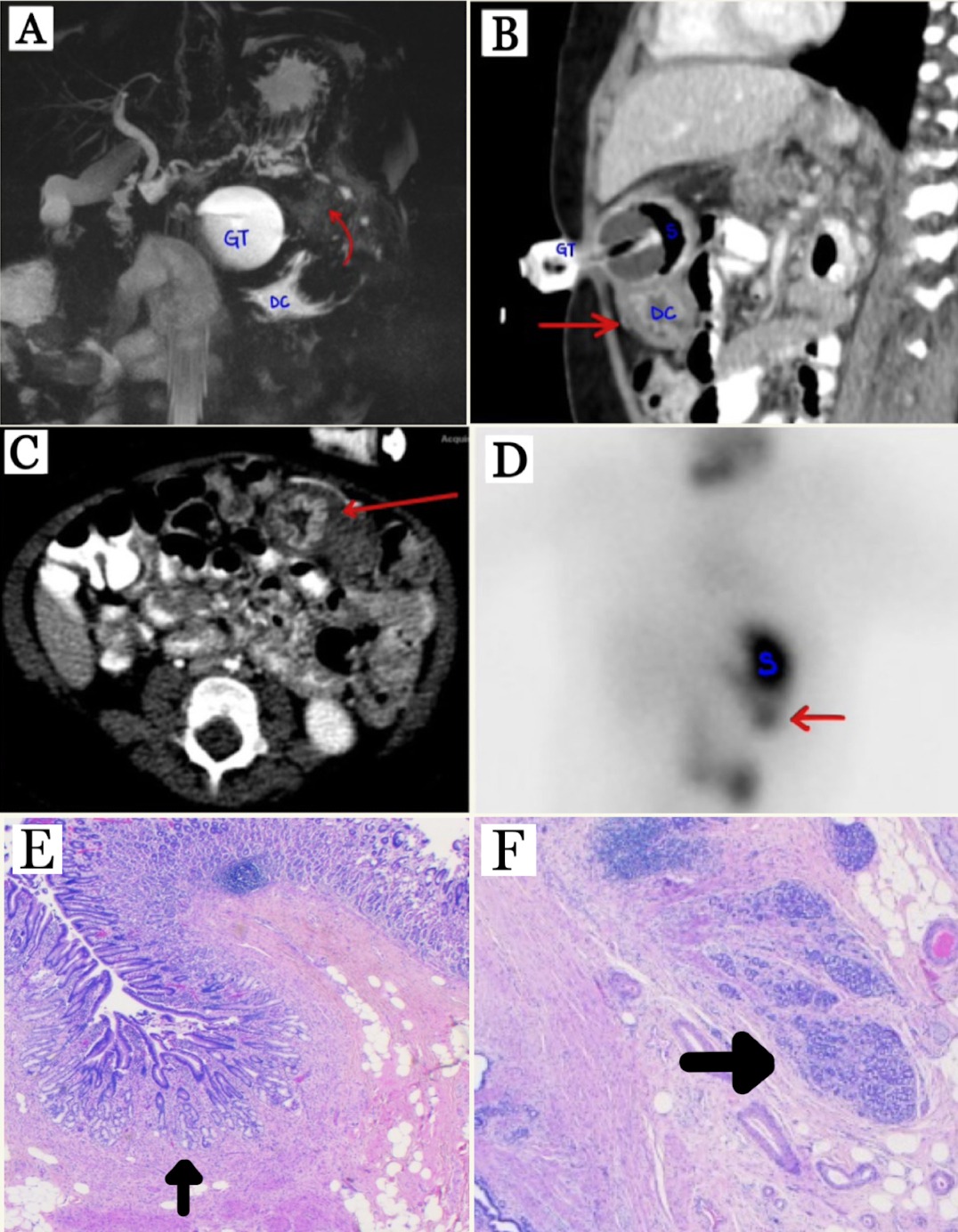
Magnetic resonance cholangiopancreatography (MRCP) finally showed signs of acute on chronic pancreatitis and an abnormal fistulous tract extending from the pancreatic tail to the greater curvature of the stomach (Figure 1. A). Computed tomography (CT) of abdomen showed a 3.7 x 2.4 x 2.3 cm thick-walled collection inferior to the gastric body with signs of gastric rugae, and confirmed the fistulous tract (Figure 1. B, C). Upper GI series did not show any communication. Subsequently, a technetium-99m scan showed uptake along the greater curvature of the stomach corresponding to focal lesion/abnormality (Figure 1. D).
The patient then underwent surgical removal of the gastric mass. The final pathology report revealed an enteric duplication cyst and incomplete annular pancreas showing gastric mucosal cells and pancreatic endocrine elements, very similar to islets of Langerhans (Figure 2).
Discussion: This is a remarkable case of pancreatic maldevelopment and enteric duplication cyst in the setting of "ring chromosome 18", a chromosomal aberration associated with a variety of phenotypical abnormalities in different organ systems. In patients with recurrent acute pancreatitis, imaging studies should be performed to exclude anatomic variations that are surgically correctable. Diagnosis requires a high index of suspicion and is often difficult because of nonspecific presenting symptoms and dependence on imaging which can be erroneous due to active inflammation in the setting of acute pancreatitis.

Disclosures:
Palack Agrawal, MD, Keshav Bhattar, MD, Claudia Rojas, MD, Diego Diaz, MD. P1478 - Enteric Duplication Cysts: An Overlooked Cause of Recurrent Pancreatitis, ACG 2024 Annual Scientific Meeting Abstracts. Philadelphia, PA: American College of Gastroenterology.
Memorial Healthcare System, Hollywood, FL
Introduction: Duplication cysts, or alimentary tract duplications, are congenital lesions of the gastrointestinal tract, with an incidence of 1 in 4500 births & a slight male preponderance. Duplication cysts of the stomach are quite rare, and most of them have been reported in children. Their presentation is diverse due to variations in location, size, presence of heterotopic mucosa, etc. which in turn makes the diagnosis challenging.
Case Description/Methods: A 2-year-old girl with history of ring chromosome 18 syndrome presented with hematemesis. Initial labs showed normal electrolytes and blood counts, elevated lipase, and mild transaminitis. A right upper quadrant ultrasound (US) revealed trace pancreatic ductal dilatation. She was treated for suspected pancreatitis but continued to have similar episodes requiring repeated hospitalisations, not responding to feed adjustments, octreotide or pancrealipase.
Magnetic resonance cholangiopancreatography (MRCP) finally showed signs of acute on chronic pancreatitis and an abnormal fistulous tract extending from the pancreatic tail to the greater curvature of the stomach (Figure 1. A). Computed tomography (CT) of abdomen showed a 3.7 x 2.4 x 2.3 cm thick-walled collection inferior to the gastric body with signs of gastric rugae, and confirmed the fistulous tract (Figure 1. B, C). Upper GI series did not show any communication. Subsequently, a technetium-99m scan showed uptake along the greater curvature of the stomach corresponding to focal lesion/abnormality (Figure 1. D).
The patient then underwent surgical removal of the gastric mass. The final pathology report revealed an enteric duplication cyst and incomplete annular pancreas showing gastric mucosal cells and pancreatic endocrine elements, very similar to islets of Langerhans (Figure 2).
Discussion: This is a remarkable case of pancreatic maldevelopment and enteric duplication cyst in the setting of "ring chromosome 18", a chromosomal aberration associated with a variety of phenotypical abnormalities in different organ systems. In patients with recurrent acute pancreatitis, imaging studies should be performed to exclude anatomic variations that are surgically correctable. Diagnosis requires a high index of suspicion and is often difficult because of nonspecific presenting symptoms and dependence on imaging which can be erroneous due to active inflammation in the setting of acute pancreatitis.

Figure: Fig (A) MRCP: Beaded appearance of pancreatic duct and fistulous tracts from the pancreatic tail to the DC measuring 2.3 x 1.9 x 2.8 cm (DC : duplication cyst)
Fig (B) CT abdomen: Gastrostomy Tube (GT) with balloon within the gastric lumen (S: stomach), DC arising from the greater curvature (red arrow)
Fig (C)Langerhansn: DC with the appearance of gastric mucosa (red arrow).
Fig (D) Technetium -99m Scan: Uptake along the greater curvature of the stomach corresponding to focal abnormality
Fig (E). Duplication cyst with gastric mucosa (black arrow)
Fig (F). Rest of pancreatic elements combining few exocrine acini with variable sized and predominant endocrine islets (black arrow), very similar to islets of Langerhans
Disclosures:
Palack Agrawal indicated no relevant financial relationships.
Keshav Bhattar indicated no relevant financial relationships.
Claudia Rojas indicated no relevant financial relationships.
Diego Diaz indicated no relevant financial relationships.
Palack Agrawal, MD, Keshav Bhattar, MD, Claudia Rojas, MD, Diego Diaz, MD. P1478 - Enteric Duplication Cysts: An Overlooked Cause of Recurrent Pancreatitis, ACG 2024 Annual Scientific Meeting Abstracts. Philadelphia, PA: American College of Gastroenterology.
