Sunday Poster Session
Category: Small Intestine
P1562 - Distal Jejunal Metastatic Melanoma Presenting as Symptomatic Anemia
Sunday, October 27, 2024
3:30 PM - 7:00 PM ET
Location: Exhibit Hall E

Has Audio

Allison Derise, MD
Louisiana State University Health
Kenner, LA
Presenting Author(s)
Allison Derise, MD1, John F. G.. Bobo, MD2, Stephen Landreneau, MD2, Jason Stibbe, MD3
1Louisiana State University Health, Kenner, LA; 2LSU, New Orleans, LA; 3LSU Health New Orleans School of Medicine, New Orleans, LA
Introduction: Metastatic melanoma disease within the GI tract is rare and most often presents when the disease is advanced causing symptoms such as bleeding, obstruction, and intussusception. Malignant melanoma is usually found within the stomach and duodenum, presents with multiple lesions, and spreads hematogenously. We present a case of biopsy-proven malignant melanoma within the distal jejunum from an unknown primary site in a patient who presented with symptomatic anemia.
Case Description/Methods: We present a 78-year-old male with history of BCC of nose, SCC of multiple sites, biopsy proven metastatic melanoma of L humerus with unknown primary s/p radiation therapy, CAD, HTN, and tobacco use who presented to the ED for hemoglobin of 5.1 from outpatient labs prior to starting immunotherapy. Per the patient, he had been feeling more fatigued with increasing DOE. He denied any signs of overt GI bleeding including hematemesis, melena, hematochezia. He has never had an EGD and his last colonoscopy 5 years ago found 5 benign polyps. He had a PET scan done a few months prior to presentation which showed a small bowel hypermetabolic focus involving a portion of the jejunum representing a segment of intussusception without CT evidence of obstruction. No definitive mass or otherwise concerning lead point was identified.
Inpatient EGD was normal, and colonoscopy had blood throughout colon and terminal ileum. This raised concern for a small bowel bleed and possible small bowel lesion considering the hypermetabolic area on the PET scan. VCE was performed and showed red blood and two mucosal deformities in the small bowel. There was mucosal edema, ulceration, discoloration, red blood, and capsule hang up near the latter lesion.
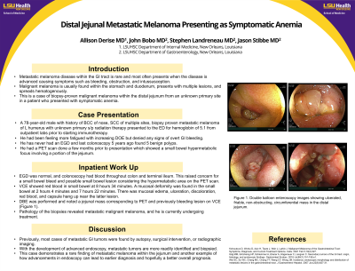
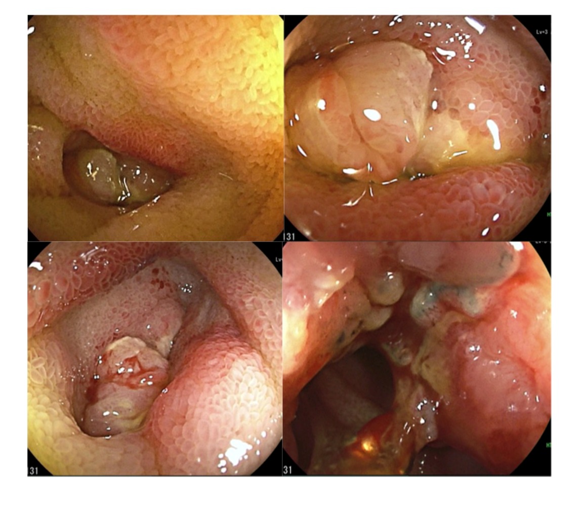
DBE was performed and noted a jejunal mass corresponding to the PET and previously bleeding lesion on VCE (Figure 1). Pathology of the biopsies revealed metastatic malignant melanoma, and he is currently undergoing further staging and treatment options.
Discussion: Previously, most cases of metastatic GI tumors were found by autopsy, surgical intervention, or radiographic imaging. With the development of advanced endoscopy such as video capsule endoscopy and single/double balloon endoscopy, metastatic tumors are more readily identified and biopsied. This case demonstrates a rare finding of metastatic melanoma within the jejunum and another example of how advancements in endoscopy can lead to earlier diagnosis and hopefully a better overall prognosis for patients.

Disclosures:
Allison Derise, MD1, John F. G.. Bobo, MD2, Stephen Landreneau, MD2, Jason Stibbe, MD3. P1562 - Distal Jejunal Metastatic Melanoma Presenting as Symptomatic Anemia, ACG 2024 Annual Scientific Meeting Abstracts. Philadelphia, PA: American College of Gastroenterology.
1Louisiana State University Health, Kenner, LA; 2LSU, New Orleans, LA; 3LSU Health New Orleans School of Medicine, New Orleans, LA
Introduction: Metastatic melanoma disease within the GI tract is rare and most often presents when the disease is advanced causing symptoms such as bleeding, obstruction, and intussusception. Malignant melanoma is usually found within the stomach and duodenum, presents with multiple lesions, and spreads hematogenously. We present a case of biopsy-proven malignant melanoma within the distal jejunum from an unknown primary site in a patient who presented with symptomatic anemia.
Case Description/Methods: We present a 78-year-old male with history of BCC of nose, SCC of multiple sites, biopsy proven metastatic melanoma of L humerus with unknown primary s/p radiation therapy, CAD, HTN, and tobacco use who presented to the ED for hemoglobin of 5.1 from outpatient labs prior to starting immunotherapy. Per the patient, he had been feeling more fatigued with increasing DOE. He denied any signs of overt GI bleeding including hematemesis, melena, hematochezia. He has never had an EGD and his last colonoscopy 5 years ago found 5 benign polyps. He had a PET scan done a few months prior to presentation which showed a small bowel hypermetabolic focus involving a portion of the jejunum representing a segment of intussusception without CT evidence of obstruction. No definitive mass or otherwise concerning lead point was identified.
Inpatient EGD was normal, and colonoscopy had blood throughout colon and terminal ileum. This raised concern for a small bowel bleed and possible small bowel lesion considering the hypermetabolic area on the PET scan. VCE was performed and showed red blood and two mucosal deformities in the small bowel. There was mucosal edema, ulceration, discoloration, red blood, and capsule hang up near the latter lesion.
DBE was performed and noted a jejunal mass corresponding to the PET and previously bleeding lesion on VCE (Figure 1). Pathology of the biopsies revealed metastatic malignant melanoma, and he is currently undergoing further staging and treatment options.
Discussion: Previously, most cases of metastatic GI tumors were found by autopsy, surgical intervention, or radiographic imaging. With the development of advanced endoscopy such as video capsule endoscopy and single/double balloon endoscopy, metastatic tumors are more readily identified and biopsied. This case demonstrates a rare finding of metastatic melanoma within the jejunum and another example of how advancements in endoscopy can lead to earlier diagnosis and hopefully a better overall prognosis for patients.

Figure: Figure 1. Double balloon enteroscopy images showing ulcerated, friable, non-obstructing, circumferential mass in the distal jejunum.
Disclosures:
Allison Derise indicated no relevant financial relationships.
John Bobo indicated no relevant financial relationships.
Stephen Landreneau indicated no relevant financial relationships.
Jason Stibbe indicated no relevant financial relationships.
Allison Derise, MD1, John F. G.. Bobo, MD2, Stephen Landreneau, MD2, Jason Stibbe, MD3. P1562 - Distal Jejunal Metastatic Melanoma Presenting as Symptomatic Anemia, ACG 2024 Annual Scientific Meeting Abstracts. Philadelphia, PA: American College of Gastroenterology.
