Sunday Poster Session
Category: Small Intestine
P1566 - Isolated Duodenal Amyloidosis Presenting as Iron Deficiency Anemia
Sunday, October 27, 2024
3:30 PM - 7:00 PM ET
Location: Exhibit Hall E

Has Audio

Pratishtha Singh, MD
New York Brooklyn Methodist Hospital
Brooklyn, NY
Presenting Author(s)
Pratishtha Singh, MD, Makda Bsrat, MD, Sara Hasan, DO, Irwin Grosman, MD
New York Brooklyn Methodist Hospital, Brooklyn, NY
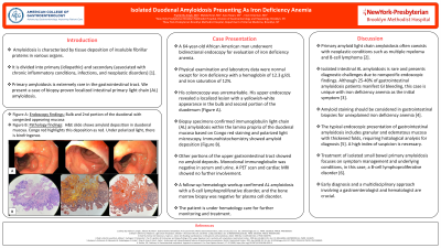
Introduction: Amyloidosis is characterized by tissue deposition of insoluble fibrillar proteins in various organs. It is divided into primary (idiopathic) and secondary (associated with chronic inflammatory conditions, infections, and neoplastic disorders). Primary amyloidosis is extremely rare in the gastrointestinal tract. We present a case of biopsy-proven localized intestinal primary light chain (AL) amyloidosis.
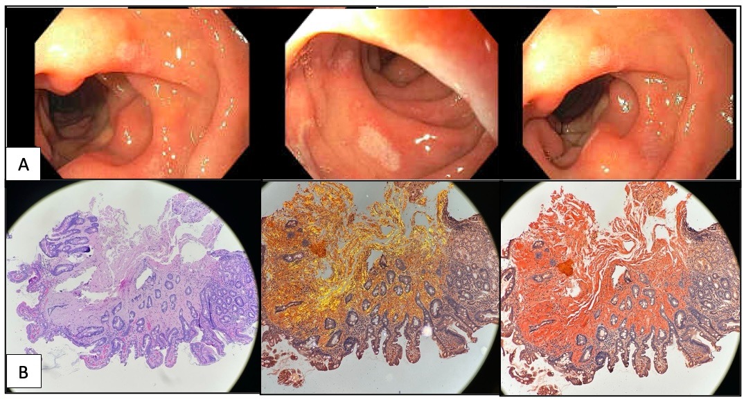
Case Description/Methods: A 64-year-old African American man underwent bidirectional endoscopy for evaluation of iron deficiency anemia. Physical examination and laboratory data were normal except for iron deficiency with a hemoglobin of 12.3 g/dL and iron saturation of 12%. His colonoscopy was unrevealing. His upper endoscopy revealed a localized lesion with a yellowish-white appearance in the bulb and second portion of the duodenum (Figure 1A). Biopsy specimens confirmed immunoglobulin light-chain (AL) amyloidosis within the lamina propria of the duodenal mucosa based on Congo red staining and polarized light microscopy. Immunohistochemistry showed amyloid deposition (Figure 1B). Other portions of the upper gastrointestinal tract showed no amyloid deposits. Monoclonal immunoglobulin was negative in serum and urine. A PET scan and cardiac MRI showed no further involvement. A follow-up hematologic workup confirmed AL amyloidosis with a B-cell lymphoproliferative disorder, and the bone marrow biopsy was negative for plasma cell disorder. The patient is under hematology care for further monitoring and treatment.
Discussion: Primary AL amyloidosis often coexists with neoplastic conditions such as multiple myeloma and B-cell lymphoma. Isolated intestinal AL amyloidosis is rare and presents diagnostic challenges due to nonspecific endoscopic findings. Although 25-40% of gastrointestinal amyloidosis patients manifest GI bleeding, this case is unique with iron deficiency anemia as the initial symptom. Amyloid staining should be considered in gastrointestinal biopsies for unexplained iron deficiency anemia. The typical endoscopic presentation of gastrointestinal amyloidosis includes granular and edematous mucosa with thickened folds, requiring histological analysis for diagnosis. A high index of suspicion is necessary. Treatment of isolated small bowel primary amyloidosis focuses on symptom management and underlying conditions, in this case, a B-cell lymphoproliferative disorder. Early diagnosis and a multidisciplinary approach involving a gastroenterologist and hematologist are crucial.

Disclosures:
Pratishtha Singh, MD, Makda Bsrat, MD, Sara Hasan, DO, Irwin Grosman, MD. P1566 - Isolated Duodenal Amyloidosis Presenting as Iron Deficiency Anemia, ACG 2024 Annual Scientific Meeting Abstracts. Philadelphia, PA: American College of Gastroenterology.
New York Brooklyn Methodist Hospital, Brooklyn, NY
Introduction: Amyloidosis is characterized by tissue deposition of insoluble fibrillar proteins in various organs. It is divided into primary (idiopathic) and secondary (associated with chronic inflammatory conditions, infections, and neoplastic disorders). Primary amyloidosis is extremely rare in the gastrointestinal tract. We present a case of biopsy-proven localized intestinal primary light chain (AL) amyloidosis.
Case Description/Methods: A 64-year-old African American man underwent bidirectional endoscopy for evaluation of iron deficiency anemia. Physical examination and laboratory data were normal except for iron deficiency with a hemoglobin of 12.3 g/dL and iron saturation of 12%. His colonoscopy was unrevealing. His upper endoscopy revealed a localized lesion with a yellowish-white appearance in the bulb and second portion of the duodenum (Figure 1A). Biopsy specimens confirmed immunoglobulin light-chain (AL) amyloidosis within the lamina propria of the duodenal mucosa based on Congo red staining and polarized light microscopy. Immunohistochemistry showed amyloid deposition (Figure 1B). Other portions of the upper gastrointestinal tract showed no amyloid deposits. Monoclonal immunoglobulin was negative in serum and urine. A PET scan and cardiac MRI showed no further involvement. A follow-up hematologic workup confirmed AL amyloidosis with a B-cell lymphoproliferative disorder, and the bone marrow biopsy was negative for plasma cell disorder. The patient is under hematology care for further monitoring and treatment.
Discussion: Primary AL amyloidosis often coexists with neoplastic conditions such as multiple myeloma and B-cell lymphoma. Isolated intestinal AL amyloidosis is rare and presents diagnostic challenges due to nonspecific endoscopic findings. Although 25-40% of gastrointestinal amyloidosis patients manifest GI bleeding, this case is unique with iron deficiency anemia as the initial symptom. Amyloid staining should be considered in gastrointestinal biopsies for unexplained iron deficiency anemia. The typical endoscopic presentation of gastrointestinal amyloidosis includes granular and edematous mucosa with thickened folds, requiring histological analysis for diagnosis. A high index of suspicion is necessary. Treatment of isolated small bowel primary amyloidosis focuses on symptom management and underlying conditions, in this case, a B-cell lymphoproliferative disorder. Early diagnosis and a multidisciplinary approach involving a gastroenterologist and hematologist are crucial.

Figure: Figure 1: A Endoscopy findings: Bulb and 2nd portion of the duodenal with congested appearing mucosa; B Pathology findings: H&E slide shows amyloid deposition in duodenal mucosa. Congo red highlights this deposition as red. Under polarized light, there is birefringence.
Disclosures:
Pratishtha Singh indicated no relevant financial relationships.
Makda Bsrat indicated no relevant financial relationships.
Sara Hasan indicated no relevant financial relationships.
Irwin Grosman indicated no relevant financial relationships.
Pratishtha Singh, MD, Makda Bsrat, MD, Sara Hasan, DO, Irwin Grosman, MD. P1566 - Isolated Duodenal Amyloidosis Presenting as Iron Deficiency Anemia, ACG 2024 Annual Scientific Meeting Abstracts. Philadelphia, PA: American College of Gastroenterology.
