Sunday Poster Session
Category: Small Intestine
P1573 - Terminal Ileum Intubation for Everyone?
Sunday, October 27, 2024
3:30 PM - 7:00 PM ET
Location: Exhibit Hall E

Has Audio

Rishika Trivedi, MD
DHR Health
Edinburg, TX
Presenting Author(s)
Rishika Trivedi, MD1, Mahmoud Barbarawi, MD2, Asif Zamir, MD, FACG3
1DHR Health, Edinburg, TX; 2DHR Health Gastroenterology, McAllen, TX; 3DHR Health Gastroenterology, Edinburg, TX
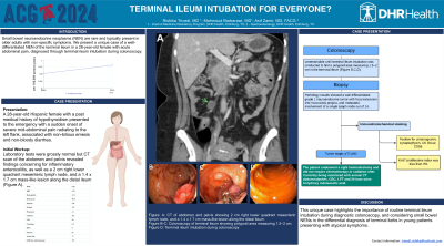
Introduction: Small bowel neuroendocrine neoplasms (NENs) are rare and typically present in older adults with non-specific symptoms. We present a unique case of a well-differentiated NEN of the terminal ileum in a 28-year-old female with acute abdominal pain, diagnosed through terminal ileum intubation during colonoscopy. This case highlights the importance of considering NENs even in young patients and routine terminal ileum intubation.
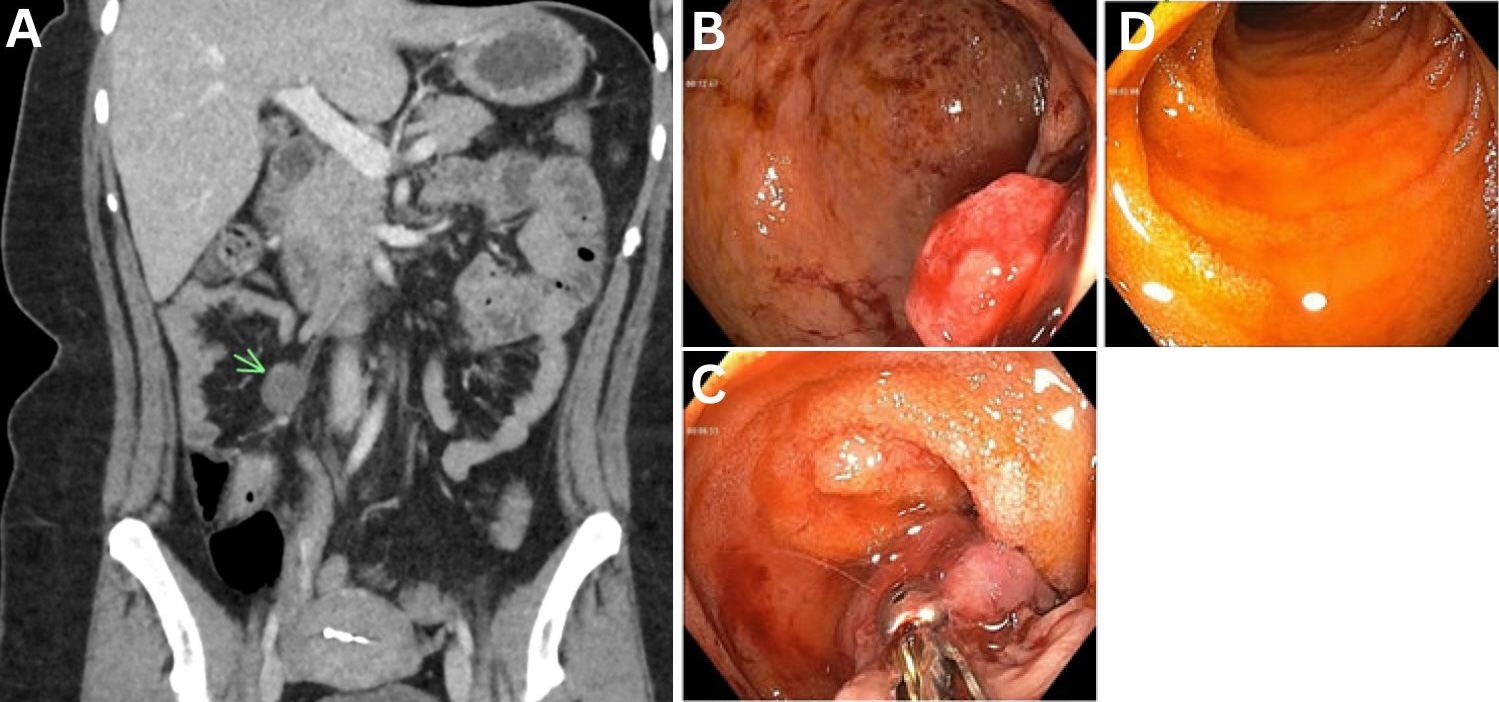
Case Description/Methods: A 28-year-old Hispanic female with a past medical history of hypothyroidism presented to the emergency with a sudden onset of severe mid-abdominal pain radiating to the left flank, associated with non-bilious emesis and non-bloody diarrhea. The initial laboratory tests were grossly normal. The CT scan of the abdomen and pelvis revealed findings concerning for inflammatory enterocolitis, as well as a 2 cm right lower quadrant mesenteric lymph node, and a 1.4 x 1.7 cm mass-like lesion along the distal ileum (Figure A). A colonoscopy was planned and upon terminal ileum intubation a polypoid area measuring 1.5–2 cm in the terminal ileum was discovered (Figure B-C). The biopsy revealed a grade 1 neuroendocrine tumor. The patient underwent a right hemicolectomy, and subsequent pathology results showed a well-differentiated neuroendocrine tumor with focal extension into muscularis propria, and metastatic involvement of a single lymph node out of 14.
The immunohistochemical staining was positive for chromogranin, synaptophysin, CK Oscar, CD56, which confirmed neuroendocrine tumor. The Ki-67 proliferative index was less than 3%. The tumor was staged as pT2 pN1. However, the patient did not require chemotherapy or radiation and is currently being monitored with annual CT abdomen/pelvis, CBC, LFT and 24 hour urine 5-Hydroxy indoleacetic acid.
Discussion: This unique case highlights the importance of routine terminal ileum intubation during diagnostic colonoscopy, and considering small bowel NETs in the differential diagnosis of terminal ileitis in young patients presenting with atypical symptoms.

Disclosures:
Rishika Trivedi, MD1, Mahmoud Barbarawi, MD2, Asif Zamir, MD, FACG3. P1573 - Terminal Ileum Intubation for Everyone?, ACG 2024 Annual Scientific Meeting Abstracts. Philadelphia, PA: American College of Gastroenterology.
1DHR Health, Edinburg, TX; 2DHR Health Gastroenterology, McAllen, TX; 3DHR Health Gastroenterology, Edinburg, TX
Introduction: Small bowel neuroendocrine neoplasms (NENs) are rare and typically present in older adults with non-specific symptoms. We present a unique case of a well-differentiated NEN of the terminal ileum in a 28-year-old female with acute abdominal pain, diagnosed through terminal ileum intubation during colonoscopy. This case highlights the importance of considering NENs even in young patients and routine terminal ileum intubation.
Case Description/Methods: A 28-year-old Hispanic female with a past medical history of hypothyroidism presented to the emergency with a sudden onset of severe mid-abdominal pain radiating to the left flank, associated with non-bilious emesis and non-bloody diarrhea. The initial laboratory tests were grossly normal. The CT scan of the abdomen and pelvis revealed findings concerning for inflammatory enterocolitis, as well as a 2 cm right lower quadrant mesenteric lymph node, and a 1.4 x 1.7 cm mass-like lesion along the distal ileum (Figure A). A colonoscopy was planned and upon terminal ileum intubation a polypoid area measuring 1.5–2 cm in the terminal ileum was discovered (Figure B-C). The biopsy revealed a grade 1 neuroendocrine tumor. The patient underwent a right hemicolectomy, and subsequent pathology results showed a well-differentiated neuroendocrine tumor with focal extension into muscularis propria, and metastatic involvement of a single lymph node out of 14.
The immunohistochemical staining was positive for chromogranin, synaptophysin, CK Oscar, CD56, which confirmed neuroendocrine tumor. The Ki-67 proliferative index was less than 3%. The tumor was staged as pT2 pN1. However, the patient did not require chemotherapy or radiation and is currently being monitored with annual CT abdomen/pelvis, CBC, LFT and 24 hour urine 5-Hydroxy indoleacetic acid.
Discussion: This unique case highlights the importance of routine terminal ileum intubation during diagnostic colonoscopy, and considering small bowel NETs in the differential diagnosis of terminal ileitis in young patients presenting with atypical symptoms.

Figure: Figure: A: CT of abdomen and pelvis showing 2 cm right lower quadrant mesenteric lymph node, and a 1.4 x 1.7 cm mass-like lesion along the distal ileum. B-C: Colonoscopy of terminal ileum showing polypoid area measuring 1.5–2 cm. D: Terminal ileum intubation during colonoscopy
Disclosures:
Rishika Trivedi indicated no relevant financial relationships.
Mahmoud Barbarawi indicated no relevant financial relationships.
Asif Zamir indicated no relevant financial relationships.
Rishika Trivedi, MD1, Mahmoud Barbarawi, MD2, Asif Zamir, MD, FACG3. P1573 - Terminal Ileum Intubation for Everyone?, ACG 2024 Annual Scientific Meeting Abstracts. Philadelphia, PA: American College of Gastroenterology.
