Sunday Poster Session
Category: Small Intestine
P1577 - A Rare Finding: Neuroendocrine Tumor Protruding Through the Ileocecal Valve
Sunday, October 27, 2024
3:30 PM - 7:00 PM ET
Location: Exhibit Hall E

Has Audio

Aya Shehata, BS
St. George's University School of Medicine
Great River, NY
Presenting Author(s)
Aya Shehata, BS1, Ahmed Shehata, MD2
1St. George's University School of Medicine, Great River, NY; 2Professional Gastroenterology Associates, Cherry Hill, NJ
Introduction: Neuroendocrine tumors (NETs) are rare neoplasms originating from neuroendocrine cells found in various organs, including the GI tract. NETs in this location are particularly intriguing due to their novelty, accounting for less than 1% of malignancies of the gastrointestinal tract. However, with this novelty comes challenges in diagnosis and management. Presenting symptoms can include intermittent bowel obstruction, changes in bowel habits, and localized pain. This case was unique in its lack of defining symptoms.
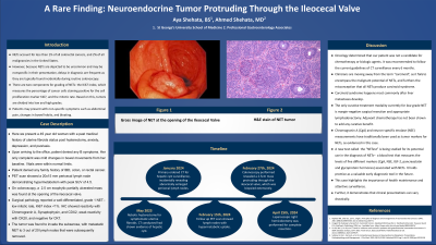
Case Description/Methods: Here we present a 49-year-old female with a past medical history of uterine fibroids status post hysterectomy after referral by oncology for colorectal cancer screening, prompted by incidental left hepatic cyst found on CT. Follow-up CT demonstrated lymphadenopathy in the ileocecal region. Patient was referred to oncology, where PET scan revealed prececal and mesenteric 20x15mm lymph nodes along the medial margin of the cecum with hypermetabolic activity, SUV 4.5. At this point, oncology referred her to our office. Upon arrival, she was asymptomatic without any abdominal pain, nausea, vomiting, B symptoms, melena, or hematochezia. On colonoscopy, we found a 3x3cm firm vascular mass originating from the opening of the ileocecal valve. Biopsy showed neuroendocrine tumor grade 1. Patient was then referred to colorectal surgery where a hand-assisted laparoscopic right hemicolectomy was performed. Surgical pathology showed tumor invasion through the muscularis propria into subserosal tissue without penetration of the overlying serosa. Proximal ileal, distal colonic, and mesenteric margins were uninvolved by the tumor, appendix showed no pathologic change. There was metastatic neuroendocrine tumor to 3 of 20 lymph nodes, stage pT3N2. IHC stained tumor cells were strongly positive for chromogranin, synaptophysin, and CDX2. The Ki67 proliferative index was less than 1%. These findings are compatible with a WHO grade 1 neuroendocrine tumor. Since then the patient is doing well. She was seen by her oncologist, who referred her to a neuroendocrine clinic for PET scan in 3 months, and surveillance CT scan every six months. No chemotherapy required.
Discussion: This rare presentation of a neuroendocrine tumor protruding through the ileocecal valve further elucidates the significance of screening and early detection. Furthermore, it bolsters the importance of a multidisciplinary approach to medicine, imaging techniques, and therapeutic strategies.

Disclosures:
Aya Shehata, BS1, Ahmed Shehata, MD2. P1577 - A Rare Finding: Neuroendocrine Tumor Protruding Through the Ileocecal Valve, ACG 2024 Annual Scientific Meeting Abstracts. Philadelphia, PA: American College of Gastroenterology.
1St. George's University School of Medicine, Great River, NY; 2Professional Gastroenterology Associates, Cherry Hill, NJ
Introduction: Neuroendocrine tumors (NETs) are rare neoplasms originating from neuroendocrine cells found in various organs, including the GI tract. NETs in this location are particularly intriguing due to their novelty, accounting for less than 1% of malignancies of the gastrointestinal tract. However, with this novelty comes challenges in diagnosis and management. Presenting symptoms can include intermittent bowel obstruction, changes in bowel habits, and localized pain. This case was unique in its lack of defining symptoms.
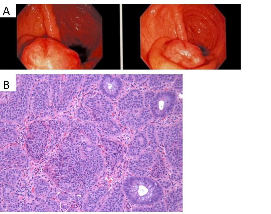
Case Description/Methods: Here we present a 49-year-old female with a past medical history of uterine fibroids status post hysterectomy after referral by oncology for colorectal cancer screening, prompted by incidental left hepatic cyst found on CT. Follow-up CT demonstrated lymphadenopathy in the ileocecal region. Patient was referred to oncology, where PET scan revealed prececal and mesenteric 20x15mm lymph nodes along the medial margin of the cecum with hypermetabolic activity, SUV 4.5. At this point, oncology referred her to our office. Upon arrival, she was asymptomatic without any abdominal pain, nausea, vomiting, B symptoms, melena, or hematochezia. On colonoscopy, we found a 3x3cm firm vascular mass originating from the opening of the ileocecal valve. Biopsy showed neuroendocrine tumor grade 1. Patient was then referred to colorectal surgery where a hand-assisted laparoscopic right hemicolectomy was performed. Surgical pathology showed tumor invasion through the muscularis propria into subserosal tissue without penetration of the overlying serosa. Proximal ileal, distal colonic, and mesenteric margins were uninvolved by the tumor, appendix showed no pathologic change. There was metastatic neuroendocrine tumor to 3 of 20 lymph nodes, stage pT3N2. IHC stained tumor cells were strongly positive for chromogranin, synaptophysin, and CDX2. The Ki67 proliferative index was less than 1%. These findings are compatible with a WHO grade 1 neuroendocrine tumor. Since then the patient is doing well. She was seen by her oncologist, who referred her to a neuroendocrine clinic for PET scan in 3 months, and surveillance CT scan every six months. No chemotherapy required.
Discussion: This rare presentation of a neuroendocrine tumor protruding through the ileocecal valve further elucidates the significance of screening and early detection. Furthermore, it bolsters the importance of a multidisciplinary approach to medicine, imaging techniques, and therapeutic strategies.

Figure: A: Gross image of NET at the opening of the Ileocecal Valve
B: H&E Stain of NET
B: H&E Stain of NET
Disclosures:
Aya Shehata indicated no relevant financial relationships.
Ahmed Shehata indicated no relevant financial relationships.
Aya Shehata, BS1, Ahmed Shehata, MD2. P1577 - A Rare Finding: Neuroendocrine Tumor Protruding Through the Ileocecal Valve, ACG 2024 Annual Scientific Meeting Abstracts. Philadelphia, PA: American College of Gastroenterology.
