Sunday Poster Session
Category: Stomach
P1635 - Granulomatous Gastritis: Another Unusual Suspect in Gastric Biopsy Leading to an Unusual Diagnostic Challenge
Sunday, October 27, 2024
3:30 PM - 7:00 PM ET
Location: Exhibit Hall E

Has Audio

Yasir Ahmed, MD
United Health Services
Binghamton, NY
Presenting Author(s)
Yasir Ahmed, MD1, Hammad Qadri, DO2, Ibrar Atiq, MD2, Ali Marhaba, MD2, Leslie Bank, MD2, Amine Hila, MD3
1United Health Services, Binghamton, NY; 2United Health Services, Wilson Medical Center, Binghamton, NY; 3UHS, Binghamton, NY
Introduction: Granulomatous gastritis (GG) is a very rare subtype of chronic gastritis that accounts for only 0.08 – 0.35% of all gastric biopsy samples. It is characterized by granulomas within the gastric mucosa or submucosa. We present a case of GG in an elderly male that led to additional workup for etiologies, once GG was identified on gastric biopsy.
Case Description/Methods:
A 67-year-old male with hypertension, diabetes mellitus type II, chronic obstructive pulmonary disease, and bipolar disorder presented with epigastric pain, loss of appetite and intermittent diarrhea. Initial laboratory workup showed mildly elevated liver associated enzymes (LAEs) and microcytic anaemia. Stool antigen testing for Helicobacter pylori (H. pylori) was negative. Computed tomography scan of the abdomen and pelvis with intravenous contrast showed fatty liver, and positive celiac and periaortic lymphadenopathy measuring up to 21.4 mm.
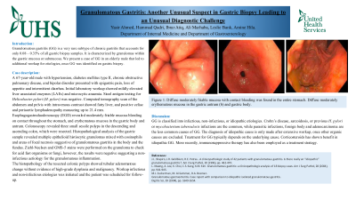
Esophagogastroduodenoscopy (EGD) revealed moderately friable mucosa bleeding on contact throughout the stomach, and erythematous mucosa in the gastric body and antrum. Colonoscopy revealed three small sessile polyps in the descending and ascending colon, which were resected. Histopathological analysis of the gastric sample revealed multiple epithelioid histiocytic granulomas mixed with eosinophils and areas of focal necrosis suggestive of granulomatous gastritis in the body and the fundus. Ziehl-Neelsen and GMS-5 stains were performed on the granuloma to check for acid fast organisms or fungi, however, the results were negative suggesting a non-infectious aetiology for the granulomatous inflammation.
The histopathology of the resected colonic polyps showed tubular adenomatous change without evidence of high-grade dysplasia and malignancy. Workup infectious and non-infectious etiologies was initiated and the patient was scheduled for follow-up.
Discussion: GG is classified into infectious, non-infectious, or idiopathic etiologies. Crohn’s disease, sarcoidosis, or previous H. pylori or mycobacterium tuberculosis infections are the common, while parasitic infections, foreign body and adenocarcinoma are the less common causes of GG. The diagnosis of idiopathic cause is only made after extensive workup, once other organic causes are excluded. Treatment for GG typically depends on the underlying cause. Corticosteroids has shown benefit in idiopathic GG. More recently, immunosuppressive therapy has also been employed as a treatment strategy.
Disclosures:
Yasir Ahmed, MD1, Hammad Qadri, DO2, Ibrar Atiq, MD2, Ali Marhaba, MD2, Leslie Bank, MD2, Amine Hila, MD3. P1635 - Granulomatous Gastritis: Another Unusual Suspect in Gastric Biopsy Leading to an Unusual Diagnostic Challenge, ACG 2024 Annual Scientific Meeting Abstracts. Philadelphia, PA: American College of Gastroenterology.
1United Health Services, Binghamton, NY; 2United Health Services, Wilson Medical Center, Binghamton, NY; 3UHS, Binghamton, NY
Introduction: Granulomatous gastritis (GG) is a very rare subtype of chronic gastritis that accounts for only 0.08 – 0.35% of all gastric biopsy samples. It is characterized by granulomas within the gastric mucosa or submucosa. We present a case of GG in an elderly male that led to additional workup for etiologies, once GG was identified on gastric biopsy.
Case Description/Methods:
A 67-year-old male with hypertension, diabetes mellitus type II, chronic obstructive pulmonary disease, and bipolar disorder presented with epigastric pain, loss of appetite and intermittent diarrhea. Initial laboratory workup showed mildly elevated liver associated enzymes (LAEs) and microcytic anaemia. Stool antigen testing for Helicobacter pylori (H. pylori) was negative. Computed tomography scan of the abdomen and pelvis with intravenous contrast showed fatty liver, and positive celiac and periaortic lymphadenopathy measuring up to 21.4 mm.
Esophagogastroduodenoscopy (EGD) revealed moderately friable mucosa bleeding on contact throughout the stomach, and erythematous mucosa in the gastric body and antrum. Colonoscopy revealed three small sessile polyps in the descending and ascending colon, which were resected. Histopathological analysis of the gastric sample revealed multiple epithelioid histiocytic granulomas mixed with eosinophils and areas of focal necrosis suggestive of granulomatous gastritis in the body and the fundus. Ziehl-Neelsen and GMS-5 stains were performed on the granuloma to check for acid fast organisms or fungi, however, the results were negative suggesting a non-infectious aetiology for the granulomatous inflammation.
The histopathology of the resected colonic polyps showed tubular adenomatous change without evidence of high-grade dysplasia and malignancy. Workup infectious and non-infectious etiologies was initiated and the patient was scheduled for follow-up.
Discussion: GG is classified into infectious, non-infectious, or idiopathic etiologies. Crohn’s disease, sarcoidosis, or previous H. pylori or mycobacterium tuberculosis infections are the common, while parasitic infections, foreign body and adenocarcinoma are the less common causes of GG. The diagnosis of idiopathic cause is only made after extensive workup, once other organic causes are excluded. Treatment for GG typically depends on the underlying cause. Corticosteroids has shown benefit in idiopathic GG. More recently, immunosuppressive therapy has also been employed as a treatment strategy.
Disclosures:
Yasir Ahmed indicated no relevant financial relationships.
Hammad Qadri indicated no relevant financial relationships.
Ibrar Atiq indicated no relevant financial relationships.
Ali Marhaba indicated no relevant financial relationships.
Leslie Bank indicated no relevant financial relationships.
Amine Hila indicated no relevant financial relationships.
Yasir Ahmed, MD1, Hammad Qadri, DO2, Ibrar Atiq, MD2, Ali Marhaba, MD2, Leslie Bank, MD2, Amine Hila, MD3. P1635 - Granulomatous Gastritis: Another Unusual Suspect in Gastric Biopsy Leading to an Unusual Diagnostic Challenge, ACG 2024 Annual Scientific Meeting Abstracts. Philadelphia, PA: American College of Gastroenterology.
