Sunday Poster Session
Category: Stomach
P1641 - Recurrent Gastric Adenocarcinoma Presenting as Pseudo-Achalasia
Sunday, October 27, 2024
3:30 PM - 7:00 PM ET
Location: Exhibit Hall E

Has Audio
- DA
Doaa Alkhader, MD
Cleveland Clinic
Abu Dhabi, Abu Dhabi, United Arab Emirates
Presenting Author(s)
Doaa Alkhader, MD, Fatma Mahmoud, MBBCh, Maryam Salma. Babar, MD, Osama Yousef, MD
Cleveland Clinic, Abu Dhabi, Abu Dhabi, United Arab Emirates
Introduction: The incidence of gastric cancer varies with high incidence in Eastern Asia. Gastric cancers are classified anatomically histologically. Endoscopy and endoscopic ultrasound (EUS) are important in the diagnosis, staging, and treatment. Management is guided by staging and includes endoscopic resection, surgery and systemic therapy.
Complete resection with negative margins is the standard goal.
We present a case with recurrent gastric adenocarcinoma presenting with pseudo-achalasia despite extensive negative workup
Case Description/Methods: 70 years old female with history of locally advanced gastric cancer post subtotal gastrectomy and chemotherapy, in complete remission on surveillance Esophagogastroduodenoscopy (EGD) and positron emission tomography–computed tomography (PET-CT), presented with epigastric pain and dysphagia. CT abdomen and EGD demonstrated esophageal distension. Barium esophagogram showed distal esophageal narrowing and proximal esophageal dilation.
EUS showed distal esophageal narrowing that was dilated and three benign reactive paraesophageal lymph nodes.
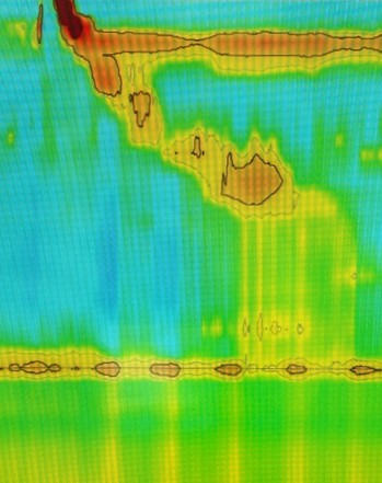
High resolution Esophageal manometry (Figure.1) displayed esophagogastric junction (EGJ) outflow obstruction. Repeat PET/CT ruled out recurrence.
Peroral Endoscopic Myotomy attempted without success. Diagnostic laparoscopy showed small bowel mesenteric carcinomatosis with tumor invasion. Frozen section confirmed recurrent gastric cancer with extensive abdominal carcinomatosis.
She was deemed unsuitable for curative therapy, with bowel obstruction, total parenteral nutrition
Discussion: 60%−70% of gastric cancer recur in 2 years with 60-70% associated mortality.3 . Recurrence patterns include loco-regional (32.4%), distant metastasis (44.3%) and peritoneal implanting (13.7%). Female patients and signet ring cell cancer are risks for peritoneal recurrence.5
In peritoneal carcinomatosis, CT compared to PET/CT had a sensitivity of 96% vs 50%, respectively.
Pseudoachalasia can be a manifestation of several pathologies with 40% due to gastro-esophageal adenocarcinoma. The proposed mechanism includes submucosal invasion with myenteric plexus destruction not identified on EGD. This was evident during our patient’s myotomy with difficulty tunneling due to severe adhesions. Pseudoachalasia may indicate underlying gastro-esophageal especially with advanced age, rapid symptom onset and EGJ outflow obstruction on manometry.
In a large series, EUS helped making the diagnosis in only two out of 18 pseudo-achalasia cases

Disclosures:
Doaa Alkhader, MD, Fatma Mahmoud, MBBCh, Maryam Salma. Babar, MD, Osama Yousef, MD. P1641 - Recurrent Gastric Adenocarcinoma Presenting as Pseudo-Achalasia, ACG 2024 Annual Scientific Meeting Abstracts. Philadelphia, PA: American College of Gastroenterology.
Cleveland Clinic, Abu Dhabi, Abu Dhabi, United Arab Emirates
Introduction: The incidence of gastric cancer varies with high incidence in Eastern Asia. Gastric cancers are classified anatomically histologically. Endoscopy and endoscopic ultrasound (EUS) are important in the diagnosis, staging, and treatment. Management is guided by staging and includes endoscopic resection, surgery and systemic therapy.
Complete resection with negative margins is the standard goal.
We present a case with recurrent gastric adenocarcinoma presenting with pseudo-achalasia despite extensive negative workup
Case Description/Methods: 70 years old female with history of locally advanced gastric cancer post subtotal gastrectomy and chemotherapy, in complete remission on surveillance Esophagogastroduodenoscopy (EGD) and positron emission tomography–computed tomography (PET-CT), presented with epigastric pain and dysphagia. CT abdomen and EGD demonstrated esophageal distension. Barium esophagogram showed distal esophageal narrowing and proximal esophageal dilation.
EUS showed distal esophageal narrowing that was dilated and three benign reactive paraesophageal lymph nodes.
High resolution Esophageal manometry (Figure.1) displayed esophagogastric junction (EGJ) outflow obstruction. Repeat PET/CT ruled out recurrence.
Peroral Endoscopic Myotomy attempted without success. Diagnostic laparoscopy showed small bowel mesenteric carcinomatosis with tumor invasion. Frozen section confirmed recurrent gastric cancer with extensive abdominal carcinomatosis.
She was deemed unsuitable for curative therapy, with bowel obstruction, total parenteral nutrition
Discussion: 60%−70% of gastric cancer recur in 2 years with 60-70% associated mortality.3 . Recurrence patterns include loco-regional (32.4%), distant metastasis (44.3%) and peritoneal implanting (13.7%). Female patients and signet ring cell cancer are risks for peritoneal recurrence.5
In peritoneal carcinomatosis, CT compared to PET/CT had a sensitivity of 96% vs 50%, respectively.
Pseudoachalasia can be a manifestation of several pathologies with 40% due to gastro-esophageal adenocarcinoma. The proposed mechanism includes submucosal invasion with myenteric plexus destruction not identified on EGD. This was evident during our patient’s myotomy with difficulty tunneling due to severe adhesions. Pseudoachalasia may indicate underlying gastro-esophageal especially with advanced age, rapid symptom onset and EGJ outflow obstruction on manometry.
In a large series, EUS helped making the diagnosis in only two out of 18 pseudo-achalasia cases

Figure: Manometry showing elevated IRP in supine with basal LES pressure of 10 mmHg and incomplete relaxation.
Disclosures:
Doaa Alkhader indicated no relevant financial relationships.
Fatma Mahmoud indicated no relevant financial relationships.
Maryam Babar indicated no relevant financial relationships.
Osama Yousef indicated no relevant financial relationships.
Doaa Alkhader, MD, Fatma Mahmoud, MBBCh, Maryam Salma. Babar, MD, Osama Yousef, MD. P1641 - Recurrent Gastric Adenocarcinoma Presenting as Pseudo-Achalasia, ACG 2024 Annual Scientific Meeting Abstracts. Philadelphia, PA: American College of Gastroenterology.
