Sunday Poster Session
Category: Stomach
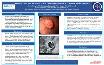
P1642 - Shedding Light on a Rare Gastric NET: Case Report on Clinical Diagnosis and Management
Sunday, October 27, 2024
3:30 PM - 7:00 PM ET
Location: Exhibit Hall E

Has Audio

Ana P. Rivera Arauz, MD
Nassau University Medical Center
Amityville, NY
Presenting Author(s)
James R. Pellegrini, MD1, Ana P. Rivera, MD2, Tulika Saggar, MD3, Atul Sinha, MD3, Sandra Gomez, MD3, Jiten Desai, MD3, Nausheer Khan, MD3
1Nassau University Medical Center, Great River, NY; 2Nassau University Medical Center, Amityville, NY; 3Nassau University Medical Center, East Meadow, NY
Introduction: Neuroendocrine tumors (NETs) of the stomach pose significant diagnostic challenges due to their diverse manifestations and potential for aggressive behavior. These tumors, representing less than 1% of all gastric neoplasms, are categorized based on their association with hypergastrinemia and conditions like atrophic gastritis and Zollinger-Ellison syndrome. This report details a case of a well-differentiated gastric NET in a 56-year-old woman with persistent gastrointestinal symptoms.
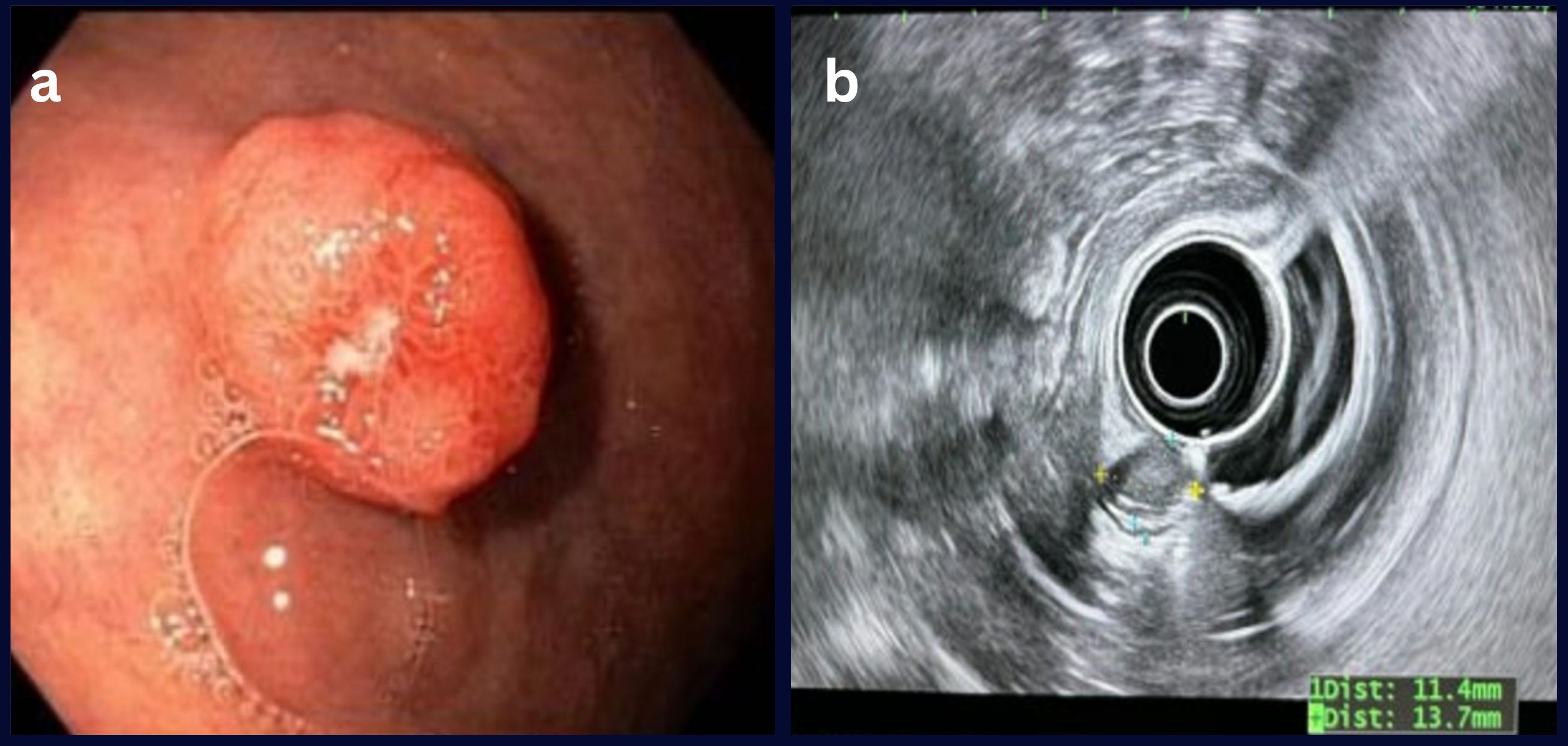
Case Description/Methods: A 56 year old female with a past medical history of hypertension, non-toxic multinodular goiter, asthma, glaucoma, and pernicious anemia presented to the GI clinic with complaints of heartburn. Gastrin level was 2471 pg/mL. The patient underwent an EGD, which revealed gastritis, a 1.5 cm sessile lesion in the body of the stomach, and multiple diminutive nodules.
Histopathology identified the larger lesion as a well-differentiated neuroendocrine tumor (NET), grade 2. The tumor had a mitotic rate of 3 per 2 mm² and a Ki-67 index between 5-10%, indicating moderate proliferative activity. Immunohistochemistry was positive for chromogranin, synaptophysin, and AE1/AE3.
Subsequently, an octreotide scan revealed no abnormal radiotracer accumulation, suggesting no metastatic spread. This finding was supported by MRIs of the chest, abdomen, and pelvis, which showed no evidence of metastasis. The patient underwent EUS to assess for depth of invasion. EUS showed the lesion extending up to the third layer. Repeat EGD was performed, and the lesion was removed via hot snare polypectomy.
The patient's management included proton pump inhibitor (PPI) therapy and close follow-up to monitor for recurrence.
Discussion: Gastric NETs require a nuanced approach to diagnosis and management due to their variable clinical presentations. The patient's elevated gastrin levels and the well-differentiated nature of the tumor suggest a Type I gastric NET, typically associated with chronic atrophic gastritis. Such tumors often have a benign course but require careful monitoring to rule out metastasis and ensure complete resection.
This case highlights the critical role of endoscopic examination and histopathological analysis in the accurate diagnosis and effective treatment of gastric NETs. Despite the rarity of gastric NETs and the variability in their presentation, thorough diagnostic evaluation and vigilant management are essential in preventing disease progression and achieving favorable outcomes.

Disclosures:
James R. Pellegrini, MD1, Ana P. Rivera, MD2, Tulika Saggar, MD3, Atul Sinha, MD3, Sandra Gomez, MD3, Jiten Desai, MD3, Nausheer Khan, MD3. P1642 - Shedding Light on a Rare Gastric NET: Case Report on Clinical Diagnosis and Management, ACG 2024 Annual Scientific Meeting Abstracts. Philadelphia, PA: American College of Gastroenterology.
1Nassau University Medical Center, Great River, NY; 2Nassau University Medical Center, Amityville, NY; 3Nassau University Medical Center, East Meadow, NY
Introduction: Neuroendocrine tumors (NETs) of the stomach pose significant diagnostic challenges due to their diverse manifestations and potential for aggressive behavior. These tumors, representing less than 1% of all gastric neoplasms, are categorized based on their association with hypergastrinemia and conditions like atrophic gastritis and Zollinger-Ellison syndrome. This report details a case of a well-differentiated gastric NET in a 56-year-old woman with persistent gastrointestinal symptoms.
Case Description/Methods: A 56 year old female with a past medical history of hypertension, non-toxic multinodular goiter, asthma, glaucoma, and pernicious anemia presented to the GI clinic with complaints of heartburn. Gastrin level was 2471 pg/mL. The patient underwent an EGD, which revealed gastritis, a 1.5 cm sessile lesion in the body of the stomach, and multiple diminutive nodules.
Histopathology identified the larger lesion as a well-differentiated neuroendocrine tumor (NET), grade 2. The tumor had a mitotic rate of 3 per 2 mm² and a Ki-67 index between 5-10%, indicating moderate proliferative activity. Immunohistochemistry was positive for chromogranin, synaptophysin, and AE1/AE3.
Subsequently, an octreotide scan revealed no abnormal radiotracer accumulation, suggesting no metastatic spread. This finding was supported by MRIs of the chest, abdomen, and pelvis, which showed no evidence of metastasis. The patient underwent EUS to assess for depth of invasion. EUS showed the lesion extending up to the third layer. Repeat EGD was performed, and the lesion was removed via hot snare polypectomy.
The patient's management included proton pump inhibitor (PPI) therapy and close follow-up to monitor for recurrence.
Discussion: Gastric NETs require a nuanced approach to diagnosis and management due to their variable clinical presentations. The patient's elevated gastrin levels and the well-differentiated nature of the tumor suggest a Type I gastric NET, typically associated with chronic atrophic gastritis. Such tumors often have a benign course but require careful monitoring to rule out metastasis and ensure complete resection.
This case highlights the critical role of endoscopic examination and histopathological analysis in the accurate diagnosis and effective treatment of gastric NETs. Despite the rarity of gastric NETs and the variability in their presentation, thorough diagnostic evaluation and vigilant management are essential in preventing disease progression and achieving favorable outcomes.

Figure: Image 1. a. EGD showing a 1.5 cm sessile gastric lesion. The Base of the lesion was injected with 1:100,000 epinephrine with adequate lift. Hot snare polypectomy was performed. b. EUS: 11mm x 13 mm sized hypoechoic gastric lesion noted on lesser curvature of gastric body in the mucosa and extending up to the third layer of gastric wall (submucosa) on radial echoendoscope. No lymphadenopathy noted.
Disclosures:
James Pellegrini indicated no relevant financial relationships.
Ana Rivera indicated no relevant financial relationships.
Tulika Saggar indicated no relevant financial relationships.
Atul Sinha indicated no relevant financial relationships.
Sandra Gomez indicated no relevant financial relationships.
Jiten Desai indicated no relevant financial relationships.
Nausheer Khan indicated no relevant financial relationships.
James R. Pellegrini, MD1, Ana P. Rivera, MD2, Tulika Saggar, MD3, Atul Sinha, MD3, Sandra Gomez, MD3, Jiten Desai, MD3, Nausheer Khan, MD3. P1642 - Shedding Light on a Rare Gastric NET: Case Report on Clinical Diagnosis and Management, ACG 2024 Annual Scientific Meeting Abstracts. Philadelphia, PA: American College of Gastroenterology.
