Sunday Poster Session
Category: Stomach
P1652 - A Case Study on Atrophic Chronic Gastritis: Dual Diagnosis and Treatment Strategies
Sunday, October 27, 2024
3:30 PM - 7:00 PM ET
Location: Exhibit Hall E

Has Audio

Jose I. De Arrigunaga, DO
University of Texas Health San Antonio
San Antonio, TX
Presenting Author(s)
Jose I.. De Arrigunaga, DO1, Umar Siddiqui, DO1, Peter Stawinski, MD2, Randy Wright, MD1
1University of Texas Health San Antonio, San Antonio, TX; 2University of Texas Health Science Center, San Antonio, TX
Introduction: There are two types of atrophic chronic gastritis (ACG): Type A gastritis (TAG) and Type B gastritis (TBG). TAG, usually found in the fundus, is caused by autoimmune conditions such as pernicious anemia (PA), where anti-parietal antibodies (APA) attack the parietal cells. TBG, typically located in the antrum, is more common and is often associated with Helicobacter pylori (H. pylori). However, increased acid production and alcohol consumption can also cause TBG. In this case, we present an elderly male with a constellation of symptoms, including weight loss, fatigue, and weakness, who was found to have features of both types of gastritis.
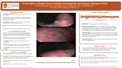
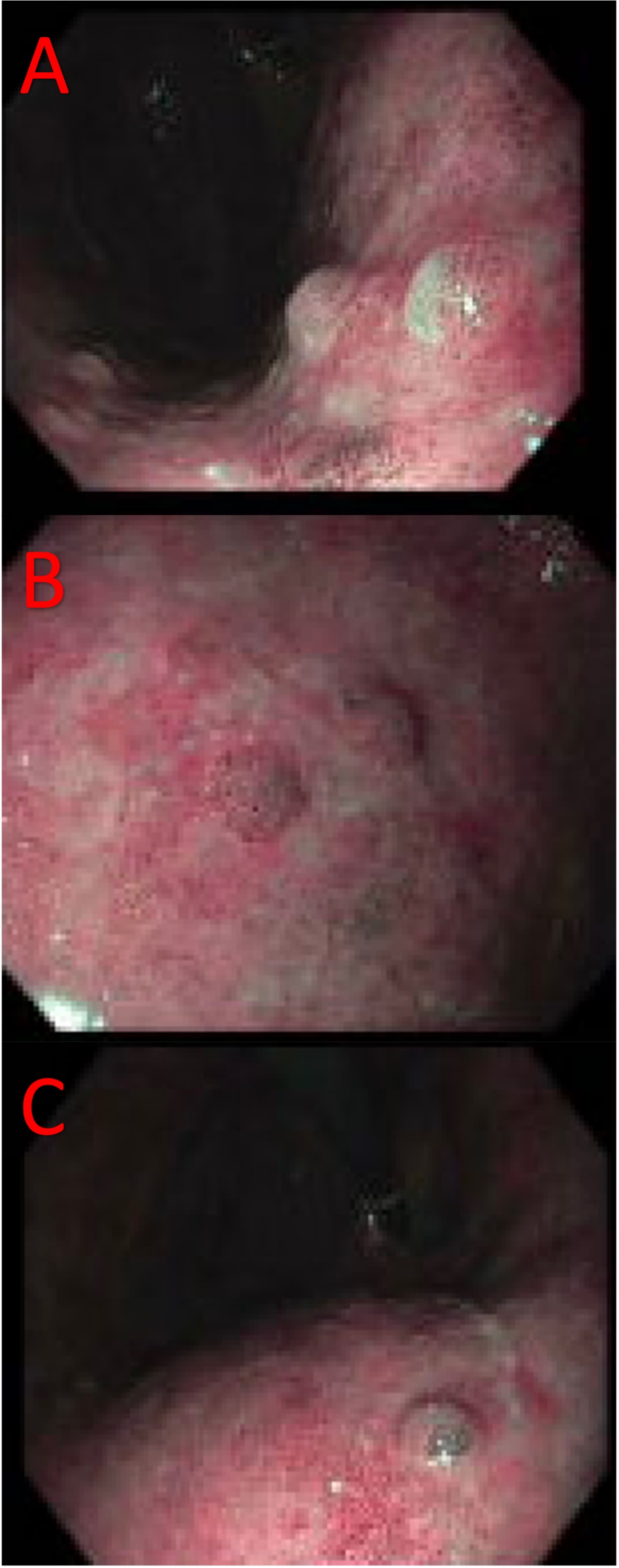
Case Description/Methods: A 71-year-old cachectic male with hypertension and hyperlipidemia presented to the hospital after three weeks of weight loss (23 pounds), fatigue, weakness, early satiety with decreased appetite, and dysphagia. Labs were significant for hemoglobin 9.7, mean corpuscular volume (MCV) 128, vitamin B12: 50, gastrin 217, positive APA, IgG 62.6, and negative intrinsic factor blocking antibody. Computed tomography of the abdomen and pelvis was negative for any intra-abdominal or pelvic abnormalities. Esophagogastroduodenoscopy (EGD) showed atrophic-appearing mucosa in the proximal stomach with a pH of 7. Additionally, multiple small gastric polyps in the proximal stomach were biopsied and showed polypoid inactive H. pylori gastritis with intestinal metaplasia. (Figure 1) The patient was started on vitamin B12 supplementation and H. pylori treatment, resulting in improvement in symptoms over the next few days.
Discussion: The two types of ACG have distinct characteristics. In this case, TAG caused by PA was the leading diagnosis due to macrocytic anemia, low vitamin B12, high gastrin, and positive APA. However, EGD revealed inactive chronic H. pylori. This finding was crucial as the treatment for TBG caused by H. pylori differs. One study demonstrated a positive association between the two types, suggesting that TAG can be caused by chronic H. pylori based on histological findings. However, this study was conducted over a prolonged period with multiple biopsies at different time intervals. Further research is needed to determine whether chronic H. pylori can cause TAG over an extended period. This case highlights the two types of ACG, their possible association, and the importance of obtaining biopsies via EGD before making a definitive diagnosis, as both vitamin B12 supplementation and H. pylori treatment were necessary for this patient.

Disclosures:
Jose I.. De Arrigunaga, DO1, Umar Siddiqui, DO1, Peter Stawinski, MD2, Randy Wright, MD1. P1652 - A Case Study on Atrophic Chronic Gastritis: Dual Diagnosis and Treatment Strategies, ACG 2024 Annual Scientific Meeting Abstracts. Philadelphia, PA: American College of Gastroenterology.
1University of Texas Health San Antonio, San Antonio, TX; 2University of Texas Health Science Center, San Antonio, TX
Introduction: There are two types of atrophic chronic gastritis (ACG): Type A gastritis (TAG) and Type B gastritis (TBG). TAG, usually found in the fundus, is caused by autoimmune conditions such as pernicious anemia (PA), where anti-parietal antibodies (APA) attack the parietal cells. TBG, typically located in the antrum, is more common and is often associated with Helicobacter pylori (H. pylori). However, increased acid production and alcohol consumption can also cause TBG. In this case, we present an elderly male with a constellation of symptoms, including weight loss, fatigue, and weakness, who was found to have features of both types of gastritis.
Case Description/Methods: A 71-year-old cachectic male with hypertension and hyperlipidemia presented to the hospital after three weeks of weight loss (23 pounds), fatigue, weakness, early satiety with decreased appetite, and dysphagia. Labs were significant for hemoglobin 9.7, mean corpuscular volume (MCV) 128, vitamin B12: 50, gastrin 217, positive APA, IgG 62.6, and negative intrinsic factor blocking antibody. Computed tomography of the abdomen and pelvis was negative for any intra-abdominal or pelvic abnormalities. Esophagogastroduodenoscopy (EGD) showed atrophic-appearing mucosa in the proximal stomach with a pH of 7. Additionally, multiple small gastric polyps in the proximal stomach were biopsied and showed polypoid inactive H. pylori gastritis with intestinal metaplasia. (Figure 1) The patient was started on vitamin B12 supplementation and H. pylori treatment, resulting in improvement in symptoms over the next few days.
Discussion: The two types of ACG have distinct characteristics. In this case, TAG caused by PA was the leading diagnosis due to macrocytic anemia, low vitamin B12, high gastrin, and positive APA. However, EGD revealed inactive chronic H. pylori. This finding was crucial as the treatment for TBG caused by H. pylori differs. One study demonstrated a positive association between the two types, suggesting that TAG can be caused by chronic H. pylori based on histological findings. However, this study was conducted over a prolonged period with multiple biopsies at different time intervals. Further research is needed to determine whether chronic H. pylori can cause TAG over an extended period. This case highlights the two types of ACG, their possible association, and the importance of obtaining biopsies via EGD before making a definitive diagnosis, as both vitamin B12 supplementation and H. pylori treatment were necessary for this patient.

Figure: Figure 1. Atrophic appearing mucosa in the proximal stomach with multiple small gastric polyps.
Disclosures:
Jose De Arrigunaga indicated no relevant financial relationships.
Umar Siddiqui indicated no relevant financial relationships.
Peter Stawinski indicated no relevant financial relationships.
Randy Wright indicated no relevant financial relationships.
Jose I.. De Arrigunaga, DO1, Umar Siddiqui, DO1, Peter Stawinski, MD2, Randy Wright, MD1. P1652 - A Case Study on Atrophic Chronic Gastritis: Dual Diagnosis and Treatment Strategies, ACG 2024 Annual Scientific Meeting Abstracts. Philadelphia, PA: American College of Gastroenterology.
