Sunday Poster Session
Category: Stomach
P1655 - Gastric Nodules as a Rare Presentation of Diffuse Large B Cell Lymphoma
Sunday, October 27, 2024
3:30 PM - 7:00 PM ET
Location: Exhibit Hall E

Has Audio

Hamzah Shariff, MD
Thomas Jefferson University Hospital
Philadelphia, PA
Presenting Author(s)
Hamzah Shariff, MD1, Ghazal Ghafari, DO, MPH2, Anila Mahesh, MD3, William Quinones, MD2, Kishore Kumar, MD4
1Thomas Jefferson University Hospital, Philadelphia, PA; 2Geisinger Health System, Danville, PA; 3Geisinger Wyoming Valley Medical Center, Wilkes-Barre, PA; 4Geisinger Community Medical Center, Scranton, PA
Introduction: Diffuse large B cell lymphoma (DLBCL) accounts for at least 30% of the non-Hodgkin lymphomas. Infiltration of the gastrointestinal (GI) tract is a well-documented manifestation in the late stage, with the stomach as a predominantly involved organ. Multiple ulcerations or polyps may be found on endoscopy, though subtle, atypical nodular appearances should not be overlooked. We present a patient found to have DLBCL with an uncommon morphology of multiple gastric nodules on endoscopy.
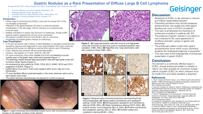
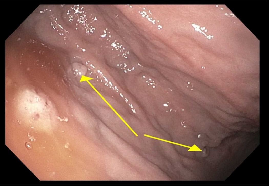
Case Description/Methods: This is a 79 year old woman with a history of atrial fibrillation on apixaban and COPD who presented with worsening dyspnea and diagnosed to have atrial fibrillation with rapid ventricular response and severe iron deficiency anemia (IDA) without overt GI bleeding. On examination the patient was afebrile and cachectic without lymphadenopathy. Laboratory values showed a hemoglobin of 7.0 g/dL, BUN 30, LDH 334. After optimization of the cardiopulmonary status, an upper endoscopy was performed. There were multiple 3 to 4 mm non-bleeding mucosal nodules seen in the gastric body which were biopsied. The pathology results showed large pleomorphic cells with high-grade nuclei and numerous mitotic figures. Immunoreactivity was positive for CD45, CD20, BCL2, MUM1, BCL6 and CD10. The findings were suggestive of DLBCL. Epstein Barre Virus and H. Pylori were negative within tumor cells via in situ hybridization. CT scan identified diffuse lymphadenopathy in the chest, abdomen, pelvis and a right hepatic dome mass. The patient declined chemotherapy and elected for comfort measures.
Discussion: Metastasis of DLBCL to the stomach is common and reflects disseminated disease. Presenting symptoms may include dyspepsia, abdominal pain, and weight loss while gastric bleeding can occur in 20-30% of patients. Further, this case re-emphasizes the importance of endoscopic evaluation in patients with IDA. The presence of gastric nodules in our patient is rare compared to the usual appearance of multifocal ulceration, polyps or gastric wall thickening. The endoscopic pattern could mimic gastric neuroendocrine tumor which occurs commonly therefore the histological diagnosis is imperative. This case owes to the heterogeneity of endoscopic presentation in gastric DLBCL.
Conclusion:
The stomach is a commonly afflicted organ in DLBCL, though endoscopic pattern is variable in nature. Our case revealed an uncommon nodular pattern of gastric DLBCL for which clinicians must be mindful of to accurately establish a diagnosis.

Disclosures:
Hamzah Shariff, MD1, Ghazal Ghafari, DO, MPH2, Anila Mahesh, MD3, William Quinones, MD2, Kishore Kumar, MD4. P1655 - Gastric Nodules as a Rare Presentation of Diffuse Large B Cell Lymphoma, ACG 2024 Annual Scientific Meeting Abstracts. Philadelphia, PA: American College of Gastroenterology.
1Thomas Jefferson University Hospital, Philadelphia, PA; 2Geisinger Health System, Danville, PA; 3Geisinger Wyoming Valley Medical Center, Wilkes-Barre, PA; 4Geisinger Community Medical Center, Scranton, PA
Introduction: Diffuse large B cell lymphoma (DLBCL) accounts for at least 30% of the non-Hodgkin lymphomas. Infiltration of the gastrointestinal (GI) tract is a well-documented manifestation in the late stage, with the stomach as a predominantly involved organ. Multiple ulcerations or polyps may be found on endoscopy, though subtle, atypical nodular appearances should not be overlooked. We present a patient found to have DLBCL with an uncommon morphology of multiple gastric nodules on endoscopy.
Case Description/Methods: This is a 79 year old woman with a history of atrial fibrillation on apixaban and COPD who presented with worsening dyspnea and diagnosed to have atrial fibrillation with rapid ventricular response and severe iron deficiency anemia (IDA) without overt GI bleeding. On examination the patient was afebrile and cachectic without lymphadenopathy. Laboratory values showed a hemoglobin of 7.0 g/dL, BUN 30, LDH 334. After optimization of the cardiopulmonary status, an upper endoscopy was performed. There were multiple 3 to 4 mm non-bleeding mucosal nodules seen in the gastric body which were biopsied. The pathology results showed large pleomorphic cells with high-grade nuclei and numerous mitotic figures. Immunoreactivity was positive for CD45, CD20, BCL2, MUM1, BCL6 and CD10. The findings were suggestive of DLBCL. Epstein Barre Virus and H. Pylori were negative within tumor cells via in situ hybridization. CT scan identified diffuse lymphadenopathy in the chest, abdomen, pelvis and a right hepatic dome mass. The patient declined chemotherapy and elected for comfort measures.
Discussion: Metastasis of DLBCL to the stomach is common and reflects disseminated disease. Presenting symptoms may include dyspepsia, abdominal pain, and weight loss while gastric bleeding can occur in 20-30% of patients. Further, this case re-emphasizes the importance of endoscopic evaluation in patients with IDA. The presence of gastric nodules in our patient is rare compared to the usual appearance of multifocal ulceration, polyps or gastric wall thickening. The endoscopic pattern could mimic gastric neuroendocrine tumor which occurs commonly therefore the histological diagnosis is imperative. This case owes to the heterogeneity of endoscopic presentation in gastric DLBCL.
Conclusion:
The stomach is a commonly afflicted organ in DLBCL, though endoscopic pattern is variable in nature. Our case revealed an uncommon nodular pattern of gastric DLBCL for which clinicians must be mindful of to accurately establish a diagnosis.

Figure: Upper endoscopy demonstrating multiple round, well circumscribed mucosal nodules on the greater curvature of the stomach
Disclosures:
Hamzah Shariff indicated no relevant financial relationships.
Ghazal Ghafari indicated no relevant financial relationships.
Anila Mahesh indicated no relevant financial relationships.
William Quinones indicated no relevant financial relationships.
Kishore Kumar indicated no relevant financial relationships.
Hamzah Shariff, MD1, Ghazal Ghafari, DO, MPH2, Anila Mahesh, MD3, William Quinones, MD2, Kishore Kumar, MD4. P1655 - Gastric Nodules as a Rare Presentation of Diffuse Large B Cell Lymphoma, ACG 2024 Annual Scientific Meeting Abstracts. Philadelphia, PA: American College of Gastroenterology.
