Sunday Poster Session
Category: Stomach
P1660 - Iron Gut
Sunday, October 27, 2024
3:30 PM - 7:00 PM ET
Location: Exhibit Hall E

- BS
Bipneet Singh, MD
Henry Ford Jackson Hospital
Jackson, MI
Presenting Author(s)
Bipneet Singh, MD1, Muhammad Zarrar Khan, MD2, Palak Patel-Rodrigues, MD3, Sruthi Ramanan, MD1, Beena Ahsan, MD3, Jason Schairer, MD3
1Henry Ford Jackson Hospital, Jackson, MI; 2Henry Ford Hospital, Royal Oak, MI; 3Henry Ford Health, Detroit, MI
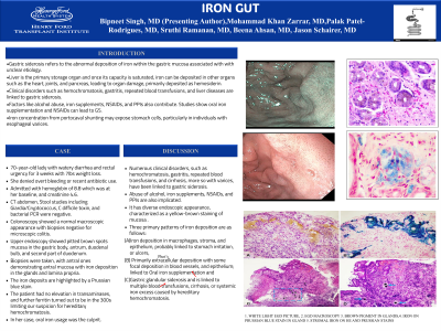
Introduction: Gastric siderosis refers to the abnormal deposition of iron within the gastric mucosa associated with with unclear etiology. Liver is the primary storage organ and once its capacity is saturated, iron can be deposited in other organs such as the heart, joints, and pancreas, leading to organ mdamage, primarily deposited as hemosiderin. Clinical disorders such as hemochromatosis, gastritis, repeated blood transfusions, and liver diseases are linked to gastric siderosis. Factors like alcohol abuse, iron supplements, NSAIDs, and PPIs also contribute. Studies show oral iron supplementation and NSAIDs can lead to GS. Iron concentration from portocaval shunting may expose stomach cells, particularly in individuals with esophageal varices.
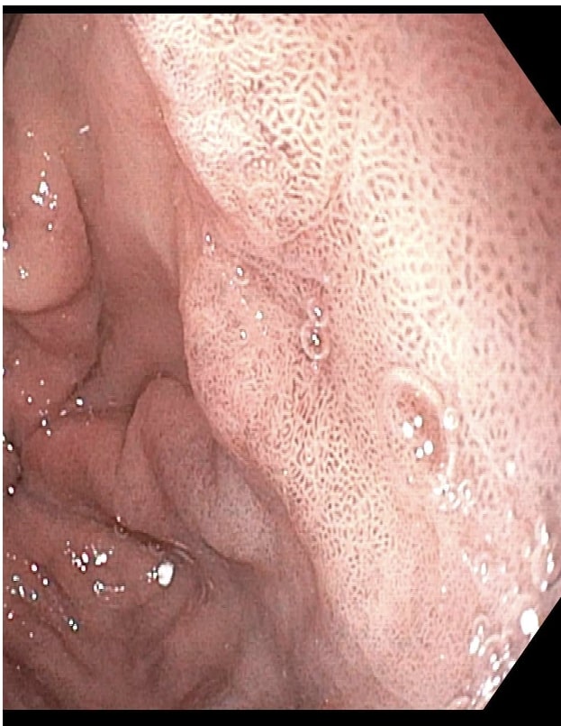
Case Description/Methods: 70-year-old lady with watery diarrhea and rectal urgency for 3 weeks with 7lbs weight loss. She denied overt bleeding, or recent antibiotic use. Admitted with hemoglobin of 8.8 which was at her baseline, and creatinine 4.6. CT abdomen, Stool studies including Giardia/Cryptococcus, C difficile toxin, and bacterial PCR were negative. Colonoscopy showed a normal macroscopic appearance with biopsies negative for microscopic colitis. Upper endoscopy showed pitted brown spots mucosa in the gastric body, antrum, duodenal bulb, and second part of duodenum. Biopsies were taken, with antral ones demonstrating antral mucosa with iron deposition in the glands and lamina propria. The iron deposits are highlighted by a Prussian blue stain. The patient had no elevation in transaminases, and further ferritin turned out to be in the 300s limiting our suspicion for hereditary hemochromatosis. In her case, oral iron usage was deemed to be the culprit for the disease.
Discussion: Numerous clinical disorders, such as hemochromatosis, gastritis, repeated blood transfusions, and cirrhosis, more so with varices, have been linked to gastric siderosis. Abuse of alcohol, iron supplements, NSAIDs, and PPIs are also implicated. It has diverse endoscopic appearance, characterized as a yellow-brown staining of mucosa . Three primary patterns of iron deposition are as follows: (A) iron deposition in macrophages, stroma, and epithelium, probably linked to stomach irritation, or ulcers, (B) primarily extracellular deposition with some focal deposition in blood vessels, and epithelium; linked to Oral iron supplementation and (C) gastric glandular siderosis and is linked to multiple blood transfusions, cirrhosis, or systemic iron excess caused by hereditary hemochromatosis.

Disclosures:
Bipneet Singh, MD1, Muhammad Zarrar Khan, MD2, Palak Patel-Rodrigues, MD3, Sruthi Ramanan, MD1, Beena Ahsan, MD3, Jason Schairer, MD3. P1660 - Iron Gut, ACG 2024 Annual Scientific Meeting Abstracts. Philadelphia, PA: American College of Gastroenterology.
1Henry Ford Jackson Hospital, Jackson, MI; 2Henry Ford Hospital, Royal Oak, MI; 3Henry Ford Health, Detroit, MI
Introduction: Gastric siderosis refers to the abnormal deposition of iron within the gastric mucosa associated with with unclear etiology. Liver is the primary storage organ and once its capacity is saturated, iron can be deposited in other organs such as the heart, joints, and pancreas, leading to organ mdamage, primarily deposited as hemosiderin. Clinical disorders such as hemochromatosis, gastritis, repeated blood transfusions, and liver diseases are linked to gastric siderosis. Factors like alcohol abuse, iron supplements, NSAIDs, and PPIs also contribute. Studies show oral iron supplementation and NSAIDs can lead to GS. Iron concentration from portocaval shunting may expose stomach cells, particularly in individuals with esophageal varices.
Case Description/Methods: 70-year-old lady with watery diarrhea and rectal urgency for 3 weeks with 7lbs weight loss. She denied overt bleeding, or recent antibiotic use. Admitted with hemoglobin of 8.8 which was at her baseline, and creatinine 4.6. CT abdomen, Stool studies including Giardia/Cryptococcus, C difficile toxin, and bacterial PCR were negative. Colonoscopy showed a normal macroscopic appearance with biopsies negative for microscopic colitis. Upper endoscopy showed pitted brown spots mucosa in the gastric body, antrum, duodenal bulb, and second part of duodenum. Biopsies were taken, with antral ones demonstrating antral mucosa with iron deposition in the glands and lamina propria. The iron deposits are highlighted by a Prussian blue stain. The patient had no elevation in transaminases, and further ferritin turned out to be in the 300s limiting our suspicion for hereditary hemochromatosis. In her case, oral iron usage was deemed to be the culprit for the disease.
Discussion: Numerous clinical disorders, such as hemochromatosis, gastritis, repeated blood transfusions, and cirrhosis, more so with varices, have been linked to gastric siderosis. Abuse of alcohol, iron supplements, NSAIDs, and PPIs are also implicated. It has diverse endoscopic appearance, characterized as a yellow-brown staining of mucosa . Three primary patterns of iron deposition are as follows: (A) iron deposition in macrophages, stroma, and epithelium, probably linked to stomach irritation, or ulcers, (B) primarily extracellular deposition with some focal deposition in blood vessels, and epithelium; linked to Oral iron supplementation and (C) gastric glandular siderosis and is linked to multiple blood transfusions, cirrhosis, or systemic iron excess caused by hereditary hemochromatosis.

Figure: Brown pigmentation of antrum
Disclosures:
Bipneet Singh indicated no relevant financial relationships.
Muhammad Zarrar Khan indicated no relevant financial relationships.
Palak Patel-Rodrigues indicated no relevant financial relationships.
Sruthi Ramanan indicated no relevant financial relationships.
Beena Ahsan indicated no relevant financial relationships.
Jason Schairer indicated no relevant financial relationships.
Bipneet Singh, MD1, Muhammad Zarrar Khan, MD2, Palak Patel-Rodrigues, MD3, Sruthi Ramanan, MD1, Beena Ahsan, MD3, Jason Schairer, MD3. P1660 - Iron Gut, ACG 2024 Annual Scientific Meeting Abstracts. Philadelphia, PA: American College of Gastroenterology.
