Sunday Poster Session
Category: Stomach
P1689 - Gastritis Cystica Profunda: A Benign Lesion That Might Not Be So Benign After All
Sunday, October 27, 2024
3:30 PM - 7:00 PM ET
Location: Exhibit Hall E

Has Audio

Kaitlyn Cronk, MD
University of Mississippi Medical Center
Jackson, MS
Presenting Author(s)
Anna Lauren G. Winter, MD, Kaitlyn Cronk, MD, Anna H.. Owings, DO, Shou-jiang Tang, MD
University of Mississippi Medical Center, Jackson, MS
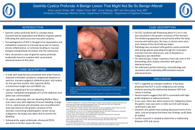
Introduction: Gastritis cystica profunda is a benign lesion characterized by hyperplasia and dilation of gastric glands infiltrating the submucosa and muscularis propria. The pathogenesis of GCP is thought to be hyperplastic and metaplastic responses to mucosal injury due to trauma, chronic inflammation, or ischemia resulting in mucosal prolapse and glandular herniation into the submucosa. Here, we present a case of gastritis cystica profunda incidentally found in a patient with concomitant adenocarcinoma of the colon.
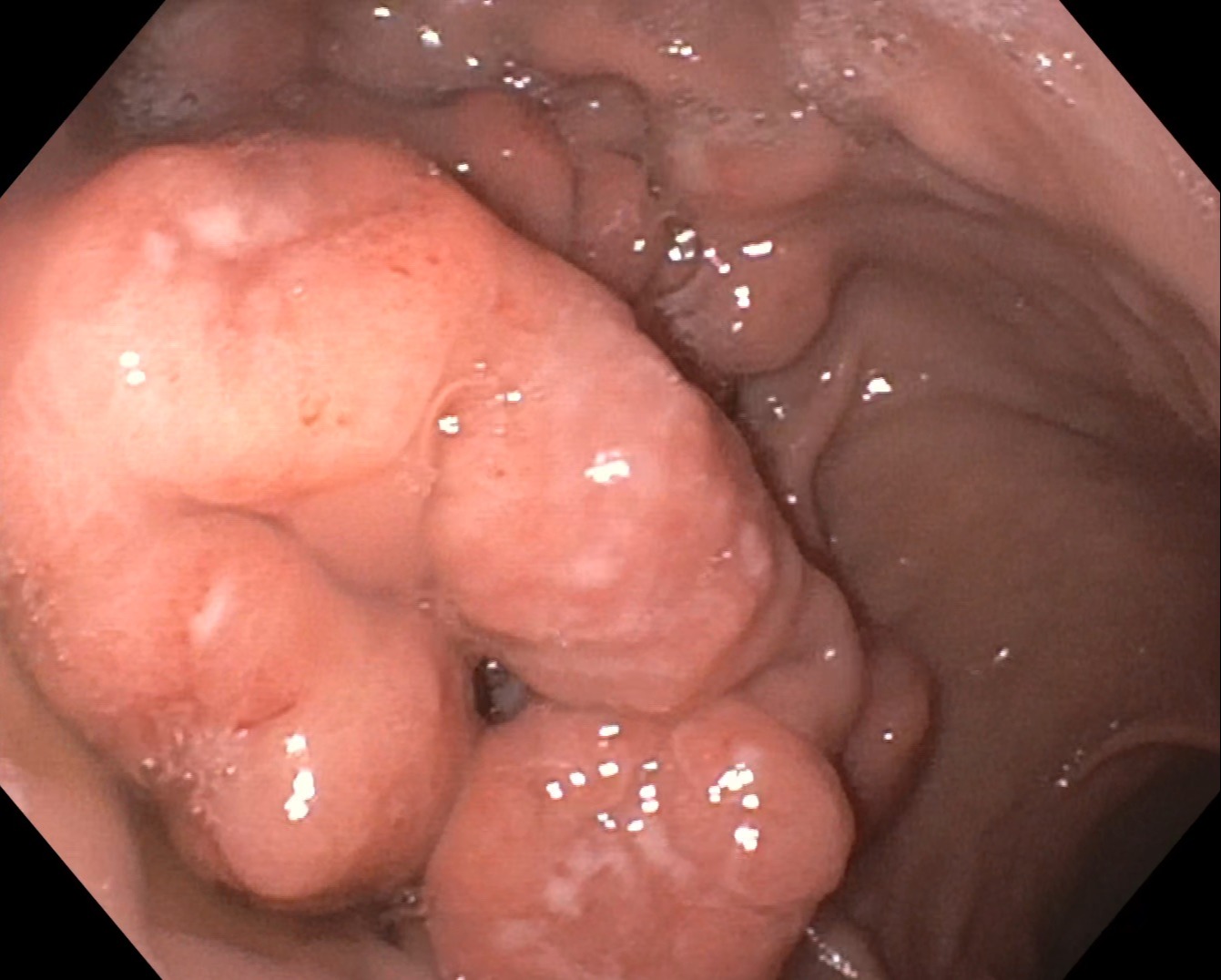
Case Description/Methods: A male with hypertension presented with ankle fracture. Reported orthostatic symptoms, progressive dyspnea on exertion, transient epigastric abdominal pain, and melena for the past few months. Also reported 30-lbs unintentional weight loss and NSAID use. Labs were significant for iron deficiency anemia. CTAP was unremarkable. On EGD, two non-bleeding Mallory Weiss tears were seen with stigmata of recent bleeding. A large 3-4 cm, submucosal and ulcerated, non-circumferential mass was found on the greater curvature of the stomach, concerning isolated gastric varices versus malignancy. No biopsy was taken due to concern for bleeding. Subsequently, upper EUS was done to further assess the mass. On EUS, localized wall thickening about 4-5 cm in size was visualized in the greater curvature of the stomach. The thickening appeared to be primarily within the deep mucosa and submucosa. No mass or varices were seen, and a biopsy of the thick fold was taken. Pathology was consistent with gastritis cystica profunda with benign glands extending through the muscularis mucosae into the submucosa, and no dysplasia or malignancy was identified. On colonoscopy, a large, suspicious mass was seen in the descending colon; biopsy consistent with gastric adenocarcinoma. He underwent partial colectomy, and pathology was consistent with moderately-differentiated invasive adenocarcinoma.
Discussion: GCP is regarded as a benign condition. It has been proposed that GCP is a pre-malignancy, but strong evidence proving the relationship between GCP and cancer is lacking. Several reports suggest that GCP is associated with high-grade dysplasia or adenocarcinoma. In our case, there was initial concern for malignancy when the gastric mass was seen on EGD, but EUS with biopsy confirmed it was GCP. However, this patient had existing adenocarcinoma of the colon, and we propose that these two findings could likely be related. Further research is needed to determine a relationship between GCP and cancer.

Disclosures:
Anna Lauren G. Winter, MD, Kaitlyn Cronk, MD, Anna H.. Owings, DO, Shou-jiang Tang, MD. P1689 - Gastritis Cystica Profunda: A Benign Lesion That Might Not Be So Benign After All, ACG 2024 Annual Scientific Meeting Abstracts. Philadelphia, PA: American College of Gastroenterology.
University of Mississippi Medical Center, Jackson, MS
Introduction: Gastritis cystica profunda is a benign lesion characterized by hyperplasia and dilation of gastric glands infiltrating the submucosa and muscularis propria. The pathogenesis of GCP is thought to be hyperplastic and metaplastic responses to mucosal injury due to trauma, chronic inflammation, or ischemia resulting in mucosal prolapse and glandular herniation into the submucosa. Here, we present a case of gastritis cystica profunda incidentally found in a patient with concomitant adenocarcinoma of the colon.
Case Description/Methods: A male with hypertension presented with ankle fracture. Reported orthostatic symptoms, progressive dyspnea on exertion, transient epigastric abdominal pain, and melena for the past few months. Also reported 30-lbs unintentional weight loss and NSAID use. Labs were significant for iron deficiency anemia. CTAP was unremarkable. On EGD, two non-bleeding Mallory Weiss tears were seen with stigmata of recent bleeding. A large 3-4 cm, submucosal and ulcerated, non-circumferential mass was found on the greater curvature of the stomach, concerning isolated gastric varices versus malignancy. No biopsy was taken due to concern for bleeding. Subsequently, upper EUS was done to further assess the mass. On EUS, localized wall thickening about 4-5 cm in size was visualized in the greater curvature of the stomach. The thickening appeared to be primarily within the deep mucosa and submucosa. No mass or varices were seen, and a biopsy of the thick fold was taken. Pathology was consistent with gastritis cystica profunda with benign glands extending through the muscularis mucosae into the submucosa, and no dysplasia or malignancy was identified. On colonoscopy, a large, suspicious mass was seen in the descending colon; biopsy consistent with gastric adenocarcinoma. He underwent partial colectomy, and pathology was consistent with moderately-differentiated invasive adenocarcinoma.
Discussion: GCP is regarded as a benign condition. It has been proposed that GCP is a pre-malignancy, but strong evidence proving the relationship between GCP and cancer is lacking. Several reports suggest that GCP is associated with high-grade dysplasia or adenocarcinoma. In our case, there was initial concern for malignancy when the gastric mass was seen on EGD, but EUS with biopsy confirmed it was GCP. However, this patient had existing adenocarcinoma of the colon, and we propose that these two findings could likely be related. Further research is needed to determine a relationship between GCP and cancer.

Figure: A large 3-4 cm, submucosal and ulcerated, non-circumferential mass was found on the greater curvature of the stomach, concerning for isolated gastric varices versus malignancy.
Disclosures:
Anna Lauren Winter indicated no relevant financial relationships.
Kaitlyn Cronk indicated no relevant financial relationships.
Anna Owings indicated no relevant financial relationships.
Shou-jiang Tang indicated no relevant financial relationships.
Anna Lauren G. Winter, MD, Kaitlyn Cronk, MD, Anna H.. Owings, DO, Shou-jiang Tang, MD. P1689 - Gastritis Cystica Profunda: A Benign Lesion That Might Not Be So Benign After All, ACG 2024 Annual Scientific Meeting Abstracts. Philadelphia, PA: American College of Gastroenterology.

