Sunday Poster Session
Category: Stomach
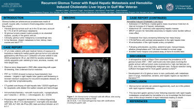
P1691 - Recurrent Glomus Tumor With Rapid Hepatic Metastasis and Hilar Lymphadenopathy in Gulf War Veteran
Sunday, October 27, 2024
3:30 PM - 7:00 PM ET
Location: Exhibit Hall E

Has Audio

Irwin A. Munoz, BS
Kirk Kerkorian School of Medicine at the University of Nevada
Presenting Author(s)
Arpinder Malhi, DO, Jasmine Dugal, DO, Amrit Narwan, MD, Irwin Munoz, BS, Preet Patel, MD, Edwin Avallone, DO
Kirk Kerkorian School of Medicine at the University of Nevada, Las Vegas, NV
Introduction: Glomus bodies are arteriovenous anastomoses made of smooth muscle cells, involved in thermoregulation via blood flow shunts. Visceral glomus tumors such as in the gastrointestinal are rare, accounting for < 1% of all GI soft tissue neoplasms. Typically, GI glomus tumors present submucosally or as ulcerated masses, usually in the gastric antrum. Clinically or histologically glomus tumor malignancy is exceedingly rare and literature has reported distant metastasis to liver, kidney, brain, ovaries and skin.
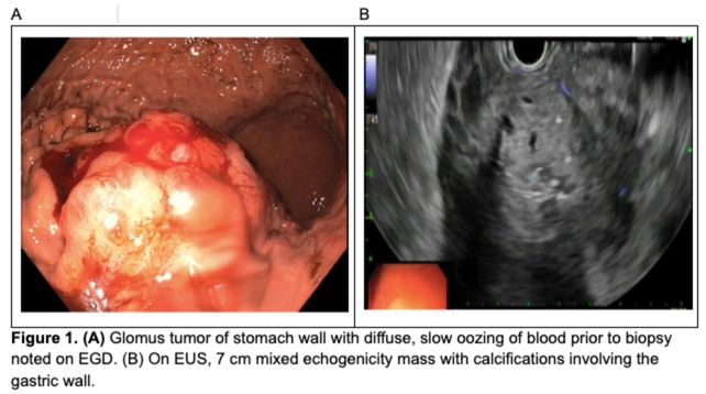
Case Description/Methods: A 46 y/o male with a history of medial gastric body wall glomus tumor diagnosed via EGD/EUS and FNA (Figure 1A/B) s/p subtotal gastrectomy, who presents for acutely worsening, epigastric abdominal pain associated with mild weight loss. Recent outpatient PET scan significant for numerous hypermetabolic liver masses, gastric wall thickening in remnant stomach suspicious for recurrent neoplasm, and new hypermetabolic right hilar lymphadenopathy. Liver biopsy one week prior was significant for neoplastic cells with dilated thin walled vessels and hemorrhage.
Physical exam remarkable for epigastric tenderness without rebound or involuntary guarding. Initial labs were significant for hemoglobin of 13.5 with an acute drop to 8.7 and elevated AST, ALT, alk phos, total and direct bilirubin. CTAP notable for gastric mass recurrence and liver metastases with numerous, solid hepatic masses noted on abdominal ultrasound. MRCP raised concern for hemobilia secondary to hepatic tumor burden. CTA abdomen concerning for active bleed within right hepatic lobe followed by subsequent IR embolization of the right hepatic artery which was actively extravasating contrast. Post-procedure, he had complete resolution of abdominal pain. He was discharged with outpatient follow-up with his community oncologist.
Discussion: The development of a GI glomus tumor is rare. A retrospective study at Mayo Clinic examined the prevalence of GI glomus tumors and found only nine cases involving the gastric antrum without lymph node involvement or high risk features on EUS, and only one histologically malignant case with surgical resection being sufficient management for gastric glomus tumors. However, this presentation of recurrent gastric glomus tumor after resection with rapid hepatic metastases complicated by hemobilia and associated with hilar lymphadenopathy highlights the possibility of aggressive nature of this tumor and its exceeding rarity compared to what is available in literature.

Disclosures:
Arpinder Malhi, DO, Jasmine Dugal, DO, Amrit Narwan, MD, Irwin Munoz, BS, Preet Patel, MD, Edwin Avallone, DO. P1691 - Recurrent Glomus Tumor With Rapid Hepatic Metastasis and Hilar Lymphadenopathy in Gulf War Veteran, ACG 2024 Annual Scientific Meeting Abstracts. Philadelphia, PA: American College of Gastroenterology.
Kirk Kerkorian School of Medicine at the University of Nevada, Las Vegas, NV
Introduction: Glomus bodies are arteriovenous anastomoses made of smooth muscle cells, involved in thermoregulation via blood flow shunts. Visceral glomus tumors such as in the gastrointestinal are rare, accounting for < 1% of all GI soft tissue neoplasms. Typically, GI glomus tumors present submucosally or as ulcerated masses, usually in the gastric antrum. Clinically or histologically glomus tumor malignancy is exceedingly rare and literature has reported distant metastasis to liver, kidney, brain, ovaries and skin.
Case Description/Methods: A 46 y/o male with a history of medial gastric body wall glomus tumor diagnosed via EGD/EUS and FNA (Figure 1A/B) s/p subtotal gastrectomy, who presents for acutely worsening, epigastric abdominal pain associated with mild weight loss. Recent outpatient PET scan significant for numerous hypermetabolic liver masses, gastric wall thickening in remnant stomach suspicious for recurrent neoplasm, and new hypermetabolic right hilar lymphadenopathy. Liver biopsy one week prior was significant for neoplastic cells with dilated thin walled vessels and hemorrhage.
Physical exam remarkable for epigastric tenderness without rebound or involuntary guarding. Initial labs were significant for hemoglobin of 13.5 with an acute drop to 8.7 and elevated AST, ALT, alk phos, total and direct bilirubin. CTAP notable for gastric mass recurrence and liver metastases with numerous, solid hepatic masses noted on abdominal ultrasound. MRCP raised concern for hemobilia secondary to hepatic tumor burden. CTA abdomen concerning for active bleed within right hepatic lobe followed by subsequent IR embolization of the right hepatic artery which was actively extravasating contrast. Post-procedure, he had complete resolution of abdominal pain. He was discharged with outpatient follow-up with his community oncologist.
Discussion: The development of a GI glomus tumor is rare. A retrospective study at Mayo Clinic examined the prevalence of GI glomus tumors and found only nine cases involving the gastric antrum without lymph node involvement or high risk features on EUS, and only one histologically malignant case with surgical resection being sufficient management for gastric glomus tumors. However, this presentation of recurrent gastric glomus tumor after resection with rapid hepatic metastases complicated by hemobilia and associated with hilar lymphadenopathy highlights the possibility of aggressive nature of this tumor and its exceeding rarity compared to what is available in literature.

Figure: Figure 1. (A) Glomus tumor of the stomach wall with diffuse, slow oozing of blood prior to biopsy noted on EGD. (B) On EUS, 7 cm mixed echogenicity mass with calcifications involving the gastric wall is noted.
Disclosures:
Arpinder Malhi indicated no relevant financial relationships.
Jasmine Dugal indicated no relevant financial relationships.
Amrit Narwan indicated no relevant financial relationships.
Irwin Munoz indicated no relevant financial relationships.
Preet Patel indicated no relevant financial relationships.
Edwin Avallone indicated no relevant financial relationships.
Arpinder Malhi, DO, Jasmine Dugal, DO, Amrit Narwan, MD, Irwin Munoz, BS, Preet Patel, MD, Edwin Avallone, DO. P1691 - Recurrent Glomus Tumor With Rapid Hepatic Metastasis and Hilar Lymphadenopathy in Gulf War Veteran, ACG 2024 Annual Scientific Meeting Abstracts. Philadelphia, PA: American College of Gastroenterology.

