Sunday Poster Session
Category: Stomach
P1693 - A Rare Gastric Glomus Tumor...Thought You Had the GIST of Gastric Masses?
Sunday, October 27, 2024
3:30 PM - 7:00 PM ET
Location: Exhibit Hall E

Has Audio
- AV
Amberly Vaughan, MD
United States Air Force
Biloxi, MS
Presenting Author(s)
Amberly Vaughan, MD, Kyle Quist, DO, Wassem Juakiem, MS, MD
United States Air Force, Biloxi, MS
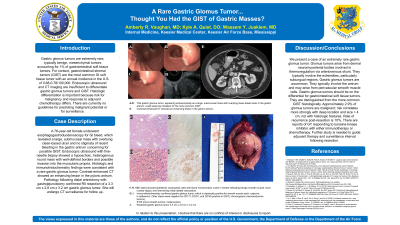
Introduction: Gastric glomus tumors (GGT) are extremely rare, typically benign, mesenchymal tumors accounting for 1% of gastrointestinal (GI) soft tissue tumors1. For context, gastrointestinal stromal tumors (GIST) are the most common GI soft tissue tumor with an annual incidence in the U.S. of 0.68-0.78/100,0002. Endoscopic ultrasound (EUS) and CT imaging are insufficient to differentiate GT and GIST3. Histologic differentiation is important because risk for malignancy and response to adjuvant chemotherapy differs. There are no guidelines for predicting malignant potential or for surveillance.
Case Description/Methods: A 76-year-old female underwent esophagogastroduodenoscopy (EGD) for GI bleed, which revealed a large, submucosal mass with overlying clean-based ulcer and no stigmata of recent bleeding in the gastric antrum concerning for possible GIST (Fig. A). EUS with fine-needle biopsy showed a hypoechoic, heterogenous round mass with well-defined borders and possible invasion into the muscularis propria. Histologic and immunohistochemistry findings were consistent with a rare gastric glomus tumor (Fig. C-F). Contrast-enhanced CT showed an enhancing lesion in the pyloric antrum (Fig. B). Pathology following distal antrectomy with gastrojejunostomy confirmed R0 resection of a 3.3 cm x 2.8 cm x 3.2 cm GGT (Fig. G). She will undergo CT surveillance.
Discussion: We present a case of an extremely rare gastric glomus tumor. Glomus tumors arise from dermal neuromyoarterial bodies involved in thermoregulation via arteriovenous shunt5. They typically involve the extremities, particularly subungual regions. Gastric GT are uncommon. They typically involve the antrum and may arise from perivascular smooth muscle cells. GGT should be on the differential for GI soft tissue tumors. They are distinguished from the more common GIST histologically. Approximately 2.9% of GT are malignant; risk correlates more strongly with deep location and size > 4 cm, not with histologic features6. Rate of recurrence post-resection is 10%4. There are reports of GT responding to tyrosine-kinase inhibitor with either immunotherapy or chemotherapy7, 8. Further study is needed to guide adjuvant therapy and surveillance interval following resection.

Disclosures:
Amberly Vaughan, MD, Kyle Quist, DO, Wassem Juakiem, MS, MD. P1693 - A Rare Gastric Glomus Tumor...Thought You Had the GIST of Gastric Masses?, ACG 2024 Annual Scientific Meeting Abstracts. Philadelphia, PA: American College of Gastroenterology.
United States Air Force, Biloxi, MS
Introduction: Gastric glomus tumors (GGT) are extremely rare, typically benign, mesenchymal tumors accounting for 1% of gastrointestinal (GI) soft tissue tumors1. For context, gastrointestinal stromal tumors (GIST) are the most common GI soft tissue tumor with an annual incidence in the U.S. of 0.68-0.78/100,0002. Endoscopic ultrasound (EUS) and CT imaging are insufficient to differentiate GT and GIST3. Histologic differentiation is important because risk for malignancy and response to adjuvant chemotherapy differs. There are no guidelines for predicting malignant potential or for surveillance.
Case Description/Methods: A 76-year-old female underwent esophagogastroduodenoscopy (EGD) for GI bleed, which revealed a large, submucosal mass with overlying clean-based ulcer and no stigmata of recent bleeding in the gastric antrum concerning for possible GIST (Fig. A). EUS with fine-needle biopsy showed a hypoechoic, heterogenous round mass with well-defined borders and possible invasion into the muscularis propria. Histologic and immunohistochemistry findings were consistent with a rare gastric glomus tumor (Fig. C-F). Contrast-enhanced CT showed an enhancing lesion in the pyloric antrum (Fig. B). Pathology following distal antrectomy with gastrojejunostomy confirmed R0 resection of a 3.3 cm x 2.8 cm x 3.2 cm GGT (Fig. G). She will undergo CT surveillance.
Discussion: We present a case of an extremely rare gastric glomus tumor. Glomus tumors arise from dermal neuromyoarterial bodies involved in thermoregulation via arteriovenous shunt5. They typically involve the extremities, particularly subungual regions. Gastric GT are uncommon. They typically involve the antrum and may arise from perivascular smooth muscle cells. GGT should be on the differential for GI soft tissue tumors. They are distinguished from the more common GIST histologically. Approximately 2.9% of GT are malignant; risk correlates more strongly with deep location and size > 4 cm, not with histologic features6. Rate of recurrence post-resection is 10%4. There are reports of GT responding to tyrosine-kinase inhibitor with either immunotherapy or chemotherapy7, 8. Further study is needed to guide adjuvant therapy and surveillance interval following resection.

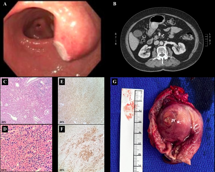
Figure: A: This gastric glomus tumor, appearing endoscopically as a large, submucosal mass with overlying clean-based ulcer in the gastric antrum, could easily be mistaken for the more common GIST.
B: Contrast-enhanced CT showed an enhancing lesion in the pyloric antrum.
C-D: H&E showed epithelioid, eosinophilic cells with bland monomorphic nuclei in sheets infiltrating benign smooth muscle, focal nuclear atypia, and intervening mildly dilated vasculature.
E-F: Immunohistochemistry stains confirmed gastric glomus tumor, which is classically positive for smooth muscle actin, calponin, h-caldesmon. Other stains were negative for CD117, DOG1, and CD34 (positive in GIST); chromogranin (neuroendocrine tumors); S100 (nerve sheath tumors, melanocytes).
G: Resected gastric glomus tumor 3.3 cm x 2.8 cm x 3.2 cm
Disclosures:
Amberly Vaughan indicated no relevant financial relationships.
Kyle Quist indicated no relevant financial relationships.
Wassem Juakiem indicated no relevant financial relationships.
Amberly Vaughan, MD, Kyle Quist, DO, Wassem Juakiem, MS, MD. P1693 - A Rare Gastric Glomus Tumor...Thought You Had the GIST of Gastric Masses?, ACG 2024 Annual Scientific Meeting Abstracts. Philadelphia, PA: American College of Gastroenterology.

