Sunday Poster Session
Category: Stomach
P1686 - Egads! That’s no EGID! A Rare Infection With Sarcina ventriculi Causing Gastric Outlet Obstruction and Eosinophilic Gastroenteritis
Sunday, October 27, 2024
3:30 PM - 7:00 PM ET
Location: Exhibit Hall E

Has Audio
- ZE
Zachary Eagle, MD
Brooke Army Medical Center
San Antonio, TX
Presenting Author(s)
Zachary Eagle, MD, Geoffrey Bader, MD
Brooke Army Medical Center, San Antonio, TX
Introduction: Sarcina Ventriculi (S ventriculi) is a Gram-positive, anaerobe with distinctive morphologic features that can survive in acidic environments. While S ventriculi is a well described pathogen in veterinary medicine, it’s pathogenicity among humans is controversial. To date, less than 100 cases have been reported, ranging from mild, non-specific symptoms to severe peritonitis, perforation, and gastric outlet obstruction (GOO). We present a rare case of GOO and suspected eosinophilic gastroenteritis (EGE) subsequently diagnosed with S ventriculi.
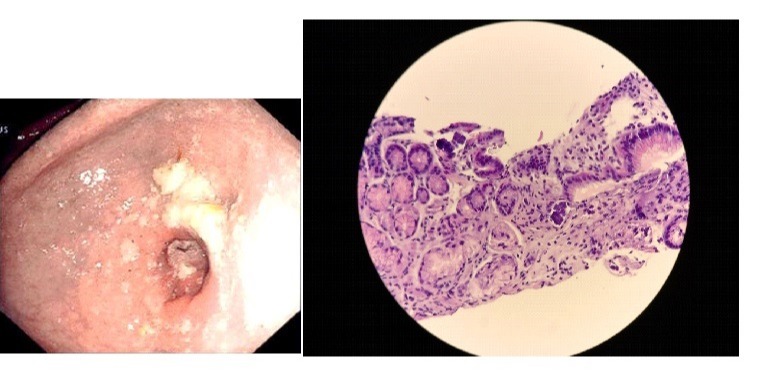
Case Description/Methods: A 28-year-old-male was admitted to the hospital for progressive abdominal pain and oral intake intolerance with emesis. Computed Tomography (CT) demonstrated hypertrophic changes involving the gastric antrum. He underwent esophagogastroduodenoscopy (EGD) which demonstrated a grossly distended stomach with non-specific erythema and a non-traversable pyloric stenosis that was successfully dilated to 10mm with through-the-scope balloon (Figure 1). Gastric biopsies demonstrated acute and chronic gastritis with prominent eosinophilic infiltrates without specified counts. He was given a diagnosis of EGE and started on esomeprazole 20mg twice daily.
When patient established care with the Lakenheath Gastroenterology service three months later, he reported minimal symptoms. Repeat EGD was performed after two week abstinence from esomeprazole and demonstrated gastric erythema, moderate volume of semi-digested food, and persistent pyloric stenosis requiring repeat dilation to 10mm. Gastric mapping biopsies of the stomach returned with chronic gastritis, up to 12 eosinophils per high power field, and organisms consistent with S ventriculi (figure 2). Patient was provided a 7-day course of ciprofloxacin and metronidazole with complete symptom resolution at follow up 3 months later. Repeat EGD with biopsy showed resolution of pyloric stenosis, chronic inactive gastritis with reduced eosinophils, and absent S ventriculi.
Discussion: S ventriculi is a poorly understood pathogen that has rarely been reported to cause catastrophic complications. The diagnosis of S ventriculi is currently dependent on histology obtained from gastric biopsies, highlighting the importance of adequate tissue sampling during EGD and discussion with pathology for potential atypical infections in obscure cases.
We hope our case brings attention to this rare clinical entity when evaluating atypical features of eosinophilic gastritis and gastric outlet obstruction.

Disclosures:
Zachary Eagle, MD, Geoffrey Bader, MD. P1686 - Egads! That’s no EGID! A Rare Infection With <i>Sarcina ventriculi</i> Causing Gastric Outlet Obstruction and Eosinophilic Gastroenteritis, ACG 2024 Annual Scientific Meeting Abstracts. Philadelphia, PA: American College of Gastroenterology.
Brooke Army Medical Center, San Antonio, TX
Introduction: Sarcina Ventriculi (S ventriculi) is a Gram-positive, anaerobe with distinctive morphologic features that can survive in acidic environments. While S ventriculi is a well described pathogen in veterinary medicine, it’s pathogenicity among humans is controversial. To date, less than 100 cases have been reported, ranging from mild, non-specific symptoms to severe peritonitis, perforation, and gastric outlet obstruction (GOO). We present a rare case of GOO and suspected eosinophilic gastroenteritis (EGE) subsequently diagnosed with S ventriculi.
Case Description/Methods: A 28-year-old-male was admitted to the hospital for progressive abdominal pain and oral intake intolerance with emesis. Computed Tomography (CT) demonstrated hypertrophic changes involving the gastric antrum. He underwent esophagogastroduodenoscopy (EGD) which demonstrated a grossly distended stomach with non-specific erythema and a non-traversable pyloric stenosis that was successfully dilated to 10mm with through-the-scope balloon (Figure 1). Gastric biopsies demonstrated acute and chronic gastritis with prominent eosinophilic infiltrates without specified counts. He was given a diagnosis of EGE and started on esomeprazole 20mg twice daily.
When patient established care with the Lakenheath Gastroenterology service three months later, he reported minimal symptoms. Repeat EGD was performed after two week abstinence from esomeprazole and demonstrated gastric erythema, moderate volume of semi-digested food, and persistent pyloric stenosis requiring repeat dilation to 10mm. Gastric mapping biopsies of the stomach returned with chronic gastritis, up to 12 eosinophils per high power field, and organisms consistent with S ventriculi (figure 2). Patient was provided a 7-day course of ciprofloxacin and metronidazole with complete symptom resolution at follow up 3 months later. Repeat EGD with biopsy showed resolution of pyloric stenosis, chronic inactive gastritis with reduced eosinophils, and absent S ventriculi.
Discussion: S ventriculi is a poorly understood pathogen that has rarely been reported to cause catastrophic complications. The diagnosis of S ventriculi is currently dependent on histology obtained from gastric biopsies, highlighting the importance of adequate tissue sampling during EGD and discussion with pathology for potential atypical infections in obscure cases.
We hope our case brings attention to this rare clinical entity when evaluating atypical features of eosinophilic gastritis and gastric outlet obstruction.

Figure: Figure 1: Image from initial upper endoscopy showing pyloric stenosis.
Figure 2: Histology slide with H&E staining that demonstrates the characteristic “cuboidal” appearance of Sarcina Ventriculi.
Figure 2: Histology slide with H&E staining that demonstrates the characteristic “cuboidal” appearance of Sarcina Ventriculi.
Disclosures:
Zachary Eagle indicated no relevant financial relationships.
Geoffrey Bader indicated no relevant financial relationships.
Zachary Eagle, MD, Geoffrey Bader, MD. P1686 - Egads! That’s no EGID! A Rare Infection With <i>Sarcina ventriculi</i> Causing Gastric Outlet Obstruction and Eosinophilic Gastroenteritis, ACG 2024 Annual Scientific Meeting Abstracts. Philadelphia, PA: American College of Gastroenterology.
