Sunday Poster Session
Category: Small Intestine
P1522 - Should We Assess Bone Density After a Celiac Disease Diagnosis in Young Adults?
Sunday, October 27, 2024
3:30 PM - 7:00 PM ET
Location: Exhibit Hall E

Has Audio

Paola M. Matos Ruiz, BS
Beth Israel Deaconess Medical Center, Harvard Medical School
Somerville, MA
Presenting Author(s)
Paola Matos Ruiz, BS1, Srihitha Akula, BS1, Jocelyn Silvester, MD, PhD2, Ciaran Kelly, MD1, Harold Rosen, MD1, Amelie Therrien, MD, MS1
1Beth Israel Deaconess Medical Center, Harvard Medical School, Boston, MA; 2Boston Children's Hospital, Boston, MA
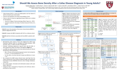
Introduction: Fracture and osteopenia are known comorbidities of celiac disease (CeD), yet there is a lack of consensus regarding indication and timing of a DEXA scan in relation to diagnosis, especially in young individuals.
Methods: Review of biopsy-proven CeD diagnosed before 65 years-old at a Celiac Center between 1/1/1999 and 12/31/2021, with a first DEXA scan after CeD diagnosis. Men < 50 years old and premenopausal women were included, and low bone density was defined as Z-score < -2.0 in this population. Clinical characteristics at the time of CeD diagnosis were reviewed and odds ratios (OR) were calculated through logistic regression for associations with low bone density. We adjusted for risk factors of low bone density, such as steroids use, family history of osteoporosis, personal history of fracture, low vitamin D levels, tobacco and heavy alcohol use.
Results: Of 907 incident CeD cases including 424 with DEXA scans, 267 met inclusion criteria. 227 DEXA scans were performed within 2 years of diagnosis. Mean bone density at L1-L4 was significantly lower compared to NHANES reference population (z-score -0.50), especially in women 20 to 40 years-old (p< 0.02). 17/227 (7.5%) had at least one z-score value < -2.0. All age groups were equally affected, but male gender (OR 3.17 95% CI 1.13-8.56) and iron deficiency anemia (IDA) at the time of CeD diagnosis (OR 3.37 95% CI 1.13-10.10) were associated with higher risk of low bone density. The 40 DEXA scans performed at first >2 years after CeD diagnosis showed similar rates of low bone density; 3/40 (7.5%) had a value < -2.0.
Logistic regression and adjusted ORs for the entire cohort supported male gender (aOR 3.36 95%CI 1.11-10.18) and being asymptomatic at the time of CeD diagnosis (aOR 4.55 95%CI 1.32–15.66), but not IDA at the time of CeD diagnosis as risk factors for low bone density.
Eleven subjects with low bone density had a second DEXA scan, on average 3.49 years after the first one, showing no difference in the bone density for each site.
Discussion: Although young women are found with a lower bone density at L1-L4 within 2 years after their diagnosis of CeD, the rate of overall low bone density defined as a z-score < -2.0 was low. This did not change according to when DEXA was first performed and did not worsen over time. Male gender and being asymptomatic at the time of CeD diagnosis were associated with a higher risk of having a low bone density in the < 50 years old men and premenopausal women population.
Disclosures:
Paola Matos Ruiz, BS1, Srihitha Akula, BS1, Jocelyn Silvester, MD, PhD2, Ciaran Kelly, MD1, Harold Rosen, MD1, Amelie Therrien, MD, MS1. P1522 - Should We Assess Bone Density After a Celiac Disease Diagnosis in Young Adults?, ACG 2024 Annual Scientific Meeting Abstracts. Philadelphia, PA: American College of Gastroenterology.
1Beth Israel Deaconess Medical Center, Harvard Medical School, Boston, MA; 2Boston Children's Hospital, Boston, MA
Introduction: Fracture and osteopenia are known comorbidities of celiac disease (CeD), yet there is a lack of consensus regarding indication and timing of a DEXA scan in relation to diagnosis, especially in young individuals.
Methods: Review of biopsy-proven CeD diagnosed before 65 years-old at a Celiac Center between 1/1/1999 and 12/31/2021, with a first DEXA scan after CeD diagnosis. Men < 50 years old and premenopausal women were included, and low bone density was defined as Z-score < -2.0 in this population. Clinical characteristics at the time of CeD diagnosis were reviewed and odds ratios (OR) were calculated through logistic regression for associations with low bone density. We adjusted for risk factors of low bone density, such as steroids use, family history of osteoporosis, personal history of fracture, low vitamin D levels, tobacco and heavy alcohol use.
Results: Of 907 incident CeD cases including 424 with DEXA scans, 267 met inclusion criteria. 227 DEXA scans were performed within 2 years of diagnosis. Mean bone density at L1-L4 was significantly lower compared to NHANES reference population (z-score -0.50), especially in women 20 to 40 years-old (p< 0.02). 17/227 (7.5%) had at least one z-score value < -2.0. All age groups were equally affected, but male gender (OR 3.17 95% CI 1.13-8.56) and iron deficiency anemia (IDA) at the time of CeD diagnosis (OR 3.37 95% CI 1.13-10.10) were associated with higher risk of low bone density. The 40 DEXA scans performed at first >2 years after CeD diagnosis showed similar rates of low bone density; 3/40 (7.5%) had a value < -2.0.
Logistic regression and adjusted ORs for the entire cohort supported male gender (aOR 3.36 95%CI 1.11-10.18) and being asymptomatic at the time of CeD diagnosis (aOR 4.55 95%CI 1.32–15.66), but not IDA at the time of CeD diagnosis as risk factors for low bone density.
Eleven subjects with low bone density had a second DEXA scan, on average 3.49 years after the first one, showing no difference in the bone density for each site.
Discussion: Although young women are found with a lower bone density at L1-L4 within 2 years after their diagnosis of CeD, the rate of overall low bone density defined as a z-score < -2.0 was low. This did not change according to when DEXA was first performed and did not worsen over time. Male gender and being asymptomatic at the time of CeD diagnosis were associated with a higher risk of having a low bone density in the < 50 years old men and premenopausal women population.
Disclosures:
Paola Matos Ruiz indicated no relevant financial relationships.
Srihitha Akula indicated no relevant financial relationships.
Jocelyn Silvester indicated no relevant financial relationships.
Ciaran Kelly indicated no relevant financial relationships.
Harold Rosen indicated no relevant financial relationships.
Amelie Therrien indicated no relevant financial relationships.
Paola Matos Ruiz, BS1, Srihitha Akula, BS1, Jocelyn Silvester, MD, PhD2, Ciaran Kelly, MD1, Harold Rosen, MD1, Amelie Therrien, MD, MS1. P1522 - Should We Assess Bone Density After a Celiac Disease Diagnosis in Young Adults?, ACG 2024 Annual Scientific Meeting Abstracts. Philadelphia, PA: American College of Gastroenterology.
