Sunday Poster Session
Category: Liver
P1351 - Benign Biliary Injury Mimicking Hepatobiliary Malignancy or Hepatobiliary Pseudotumor
Sunday, October 27, 2024
3:30 PM - 7:00 PM ET
Location: Exhibit Hall E

Has Audio

Syed Mujtaba Baqir, MD
Maimonides Medical Center
Brooklyn, NY
Presenting Author(s)
Syed Mujtaba Baqir, MD1, Sarah Meribout, MD1, Ahmed Salem, MBBCh1, Neha Sharma, MBBS, MD1, Meredith E. Pittman, MD1, Linda A. Lee, MD, MBA2
1Maimonides Medical Center, Brooklyn, NY; 2North Shore University Hospital - Northwell Health, New Hyde Park, NY
Introduction: Hepatic pseudotumors are uncommon non-neoplastic lesions that resemble neoplasia both clinically and radiologically. One cause of a hepatic pseudotumor is portal biliopathy, a type of liver injury that arises in the setting of portal cavernoma and extrahepatic portal vein obstruction of non-neoplastic and non-cirrhotic origin. Portal biliopathy can pose considerable diagnostic challenges for clinicians and pathologists. We present a case of a patient with a suspected malignant liver mass in the setting of adenopathy who was found to have pathologic changes consistent with portal biliopathy.
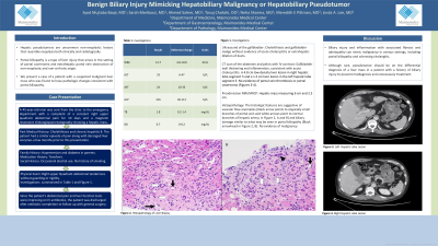
Case Description/Methods: A 45-year-old man with a history of cholelithiasis and chronic Hepatitis B was sent from the clinic to the emergency department with a complaint of a constant right upper quadrant abdominal pain for 10 days and a hepatic mass measuring 6 cm and 2.5 cm on Magnetic resonance imaging/Magnetic resonance cholangiopancreatography (MRI/MRCP). Of note, the patient had a similar episode of pain along with deranged liver enzymes a few months prior to this presentation. Initial investigation was pertinent for mild leukocytosis, mild hyperbilirubinemia, and an elevated alkaline phosphatase. Ultrasound of the gallbladder revealed cholelithiasis and gallbladder sludge without evidence of acute cholecystitis or extrahepatic dilation of ducts. CT scan of the abdomen and pelvis with IV contrast revealed gallbladder wall thickening and inflammation, consistent with acute cholecystitis. Biopsy of the largest liver lesion was consistent with biliary injury, possibly portal biliopathy. Since the patient’s abdominal pain and liver function tests were improving, the patient was discharged after antibiotic completion to follow-up with general surgery.
Discussion: Biliary injury and inflammation with associated fibrosis and adenopathy can mimic malignancy in various settings, including portal biliopathy and sclerosing cholangitis. Although rare, pseudotumor should be on the differential diagnosis of a liver mass in a patient with a history of biliary injury in order to prevent misdiagnosis and unnecessary treatment.

Disclosures:
Syed Mujtaba Baqir, MD1, Sarah Meribout, MD1, Ahmed Salem, MBBCh1, Neha Sharma, MBBS, MD1, Meredith E. Pittman, MD1, Linda A. Lee, MD, MBA2. P1351 - Benign Biliary Injury Mimicking Hepatobiliary Malignancy or Hepatobiliary Pseudotumor, ACG 2024 Annual Scientific Meeting Abstracts. Philadelphia, PA: American College of Gastroenterology.
1Maimonides Medical Center, Brooklyn, NY; 2North Shore University Hospital - Northwell Health, New Hyde Park, NY
Introduction: Hepatic pseudotumors are uncommon non-neoplastic lesions that resemble neoplasia both clinically and radiologically. One cause of a hepatic pseudotumor is portal biliopathy, a type of liver injury that arises in the setting of portal cavernoma and extrahepatic portal vein obstruction of non-neoplastic and non-cirrhotic origin. Portal biliopathy can pose considerable diagnostic challenges for clinicians and pathologists. We present a case of a patient with a suspected malignant liver mass in the setting of adenopathy who was found to have pathologic changes consistent with portal biliopathy.
Case Description/Methods: A 45-year-old man with a history of cholelithiasis and chronic Hepatitis B was sent from the clinic to the emergency department with a complaint of a constant right upper quadrant abdominal pain for 10 days and a hepatic mass measuring 6 cm and 2.5 cm on Magnetic resonance imaging/Magnetic resonance cholangiopancreatography (MRI/MRCP). Of note, the patient had a similar episode of pain along with deranged liver enzymes a few months prior to this presentation. Initial investigation was pertinent for mild leukocytosis, mild hyperbilirubinemia, and an elevated alkaline phosphatase. Ultrasound of the gallbladder revealed cholelithiasis and gallbladder sludge without evidence of acute cholecystitis or extrahepatic dilation of ducts. CT scan of the abdomen and pelvis with IV contrast revealed gallbladder wall thickening and inflammation, consistent with acute cholecystitis. Biopsy of the largest liver lesion was consistent with biliary injury, possibly portal biliopathy. Since the patient’s abdominal pain and liver function tests were improving, the patient was discharged after antibiotic completion to follow-up with general surgery.
Discussion: Biliary injury and inflammation with associated fibrosis and adenopathy can mimic malignancy in various settings, including portal biliopathy and sclerosing cholangitis. Although rare, pseudotumor should be on the differential diagnosis of a liver mass in a patient with a history of biliary injury in order to prevent misdiagnosis and unnecessary treatment.

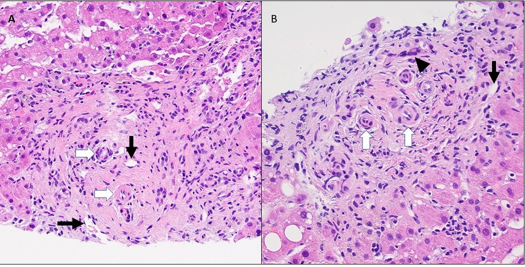
Figure: The histologic features are suggestive of vascular flow anomalies (black arrow points to atypically small branches of portal vein and white arrows point to normal branches of hepatic artery, in A and B) and biliary damage similar to what may be seen in portal biliopathy (Black arrow head in B). No evidence of malignancy.
Disclosures:
Syed Mujtaba Baqir indicated no relevant financial relationships.
Sarah Meribout indicated no relevant financial relationships.
Ahmed Salem indicated no relevant financial relationships.
Neha Sharma indicated no relevant financial relationships.
Meredith Pittman indicated no relevant financial relationships.
Linda Lee indicated no relevant financial relationships.
Syed Mujtaba Baqir, MD1, Sarah Meribout, MD1, Ahmed Salem, MBBCh1, Neha Sharma, MBBS, MD1, Meredith E. Pittman, MD1, Linda A. Lee, MD, MBA2. P1351 - Benign Biliary Injury Mimicking Hepatobiliary Malignancy or Hepatobiliary Pseudotumor, ACG 2024 Annual Scientific Meeting Abstracts. Philadelphia, PA: American College of Gastroenterology.
