Sunday Poster Session
Category: Liver
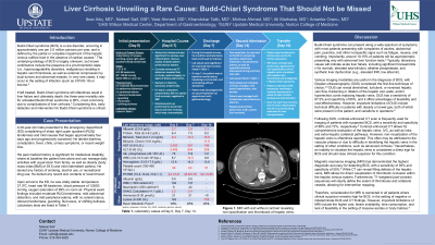
P1353 - Liver Cirrhosis Unveiling a Rare Cause: Budd-Chiari Syndrome That Should Not be Missed
Sunday, October 27, 2024
3:30 PM - 7:00 PM ET
Location: Exhibit Hall E

Has Audio

Nabeel Saif
SUNY Upstate Medical University
Syracuse, NY
Presenting Author(s)
Ibrar Atiq, MD1, Nabeel Saif, 2, Yasir Ahmed, MD1, Khandokar Talib, MD1, Minhaz Ahmad, MD1, Ali Marhaba, MD1, Amanke Oranu, MD1
1United Health Services, Wilson Medical Center, Binghamton, NY; 2SUNY Upstate Medical University, Syracuse, NY
Introduction: Budd-Chiari syndrome (BCS) is a rare disorder, If left treated, BCS will oftentimes result in liver failure and ultimately death; the 3-year mortality rate for untreated BCS is 90%, most commonly due to complications of liver cirrhosis. Considering this, early detection and intervention for BCS is critical.
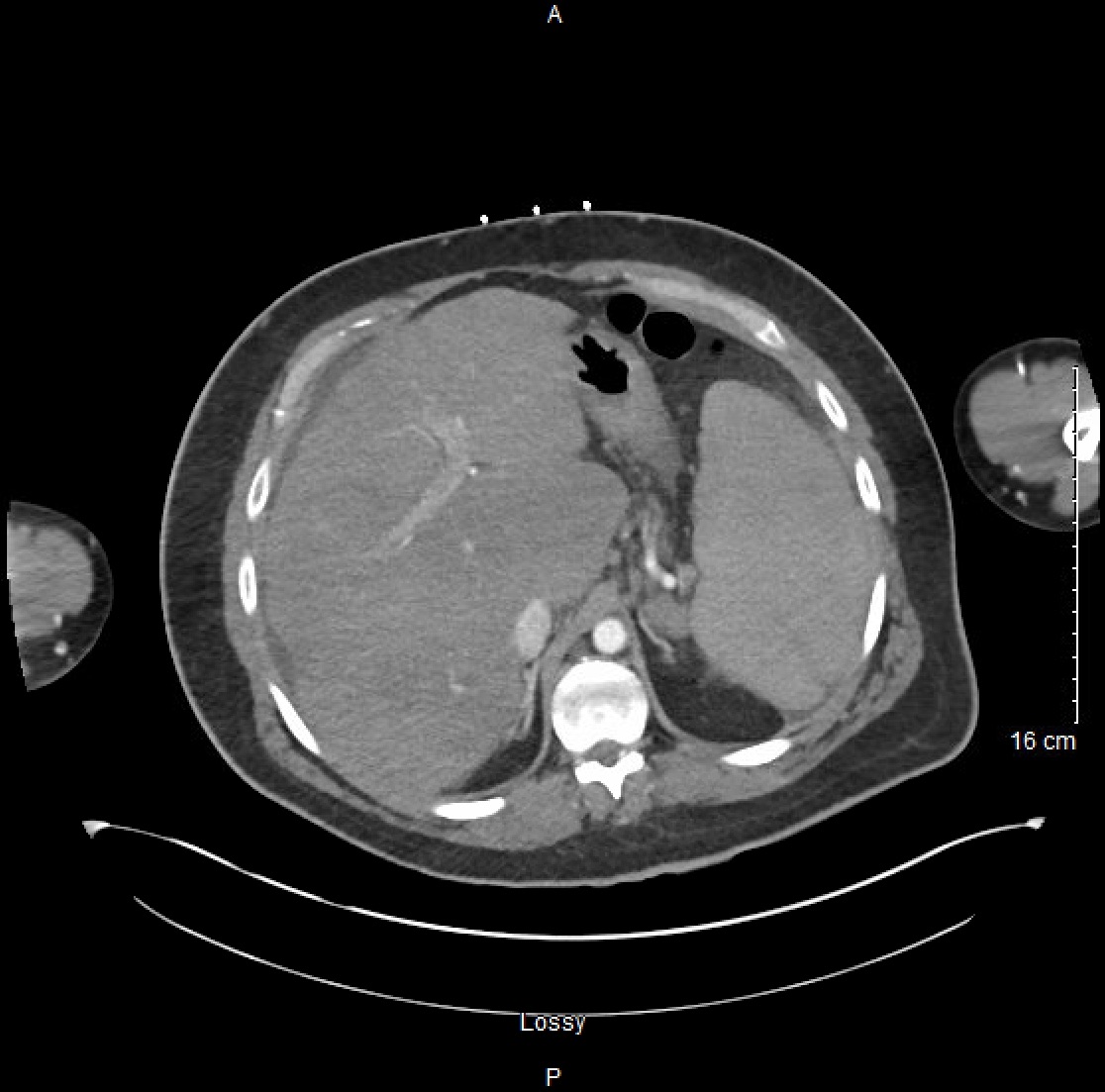
Case Description/Methods: A 29-year-old male with a medical history of intellectual disability and morbid obesity presented to the emergency (ER) complaining of a sharp right upper quadrant and mild nausea for four days. He denied any history of smoking, alcohol or recreational drug use. Upon arrival at the ER, he was vitally stable. Laboratory tests were significant for markedly elevated transaminase levels ALT and AST both >3000 U/L), INR of 3.8, platelet count 44,000/mm3, total bilirubin of 9.5 mg/dl, and alkaline phosphatase 103 U/L. Venous duplex showed a patent portal venous system. A contrast-enhanced CT of the abdomen which was significant for moderate ascites, liver cirrhosis, splenomegaly (22.2 cm), with no signs of biliary obstruction[1]. Patient underwent extensive evaluation for causes of chronic liver disease, all of which were unremarkable. Ultimately the patient was transferred to a tertiary care center for evaluation, an MRI with and without contrast of the liver was performed, which was significant for non-opacification of the hepatic veins, consistent with hepatic vein thrombosis and a diagnosis of Budd-Chiari syndrome. The following day, the patient underwent the direct intrahepatic portocaval shunt (DIPS) procedure. Following DIPS procedure, the patient’s liver function studies and total bilirubin levels markedly improved. However, over the course of the next three months, his liver function deteriorated, and he is currently being evaluated for liver transplantation.
Discussion: Various imaging modalities are useful in the diagnosis of BCS, with Doppler ultrasonography (DUS) considered the first-line followed by CT scan and MRI. However, important limitations of DUS include technical difficulty in patients with obesity or bowel gas, both of which were present in the presented patient, and variations in operative dependency. While CT findings of BCS include filling defects of the hepatic veins and/or IVC, MRI allows for direct visualization of a thrombotic occlusion within the hepatic venous system. Therefore, consideration for MRI is warranted in all patients where clinical suspicion remains high for BCS, in the setting of negative or indeterminate DUS and CT findings.

Disclosures:
Ibrar Atiq, MD1, Nabeel Saif, 2, Yasir Ahmed, MD1, Khandokar Talib, MD1, Minhaz Ahmad, MD1, Ali Marhaba, MD1, Amanke Oranu, MD1. P1353 - Liver Cirrhosis Unveiling a Rare Cause: Budd-Chiari Syndrome That Should Not be Missed, ACG 2024 Annual Scientific Meeting Abstracts. Philadelphia, PA: American College of Gastroenterology.
1United Health Services, Wilson Medical Center, Binghamton, NY; 2SUNY Upstate Medical University, Syracuse, NY
Introduction: Budd-Chiari syndrome (BCS) is a rare disorder, If left treated, BCS will oftentimes result in liver failure and ultimately death; the 3-year mortality rate for untreated BCS is 90%, most commonly due to complications of liver cirrhosis. Considering this, early detection and intervention for BCS is critical.
Case Description/Methods: A 29-year-old male with a medical history of intellectual disability and morbid obesity presented to the emergency (ER) complaining of a sharp right upper quadrant and mild nausea for four days. He denied any history of smoking, alcohol or recreational drug use. Upon arrival at the ER, he was vitally stable. Laboratory tests were significant for markedly elevated transaminase levels ALT and AST both >3000 U/L), INR of 3.8, platelet count 44,000/mm3, total bilirubin of 9.5 mg/dl, and alkaline phosphatase 103 U/L. Venous duplex showed a patent portal venous system. A contrast-enhanced CT of the abdomen which was significant for moderate ascites, liver cirrhosis, splenomegaly (22.2 cm), with no signs of biliary obstruction[1]. Patient underwent extensive evaluation for causes of chronic liver disease, all of which were unremarkable. Ultimately the patient was transferred to a tertiary care center for evaluation, an MRI with and without contrast of the liver was performed, which was significant for non-opacification of the hepatic veins, consistent with hepatic vein thrombosis and a diagnosis of Budd-Chiari syndrome. The following day, the patient underwent the direct intrahepatic portocaval shunt (DIPS) procedure. Following DIPS procedure, the patient’s liver function studies and total bilirubin levels markedly improved. However, over the course of the next three months, his liver function deteriorated, and he is currently being evaluated for liver transplantation.
Discussion: Various imaging modalities are useful in the diagnosis of BCS, with Doppler ultrasonography (DUS) considered the first-line followed by CT scan and MRI. However, important limitations of DUS include technical difficulty in patients with obesity or bowel gas, both of which were present in the presented patient, and variations in operative dependency. While CT findings of BCS include filling defects of the hepatic veins and/or IVC, MRI allows for direct visualization of a thrombotic occlusion within the hepatic venous system. Therefore, consideration for MRI is warranted in all patients where clinical suspicion remains high for BCS, in the setting of negative or indeterminate DUS and CT findings.

Figure: The liver is heterogeneous and bulky and contour consistent with cirrhosis. The spleen is enlarged, measuring 22.2 cm
Disclosures:
Ibrar Atiq indicated no relevant financial relationships.
Nabeel Saif indicated no relevant financial relationships.
Yasir Ahmed indicated no relevant financial relationships.
Khandokar Talib indicated no relevant financial relationships.
Minhaz Ahmad indicated no relevant financial relationships.
Ali Marhaba indicated no relevant financial relationships.
Amanke Oranu indicated no relevant financial relationships.
Ibrar Atiq, MD1, Nabeel Saif, 2, Yasir Ahmed, MD1, Khandokar Talib, MD1, Minhaz Ahmad, MD1, Ali Marhaba, MD1, Amanke Oranu, MD1. P1353 - Liver Cirrhosis Unveiling a Rare Cause: Budd-Chiari Syndrome That Should Not be Missed, ACG 2024 Annual Scientific Meeting Abstracts. Philadelphia, PA: American College of Gastroenterology.
