Sunday Poster Session
Category: Liver
P1355 - A Rare Case of Spontaneous Regression of Hepatocellular Carcinoma
Sunday, October 27, 2024
3:30 PM - 7:00 PM ET
Location: Exhibit Hall E

Has Audio

Colin Hartgerink, BS
Wayne State University School of Medicine
Detroit, MI
Presenting Author(s)
Award: Presidential Poster Award
Colin Hartgerink, BS1, Dilip Moonka, MD2, Syed-Mohammed Jafri, MD2
1Wayne State University School of Medicine, Detroit, MI; 2Henry Ford Health, Detroit, MI
Introduction: Spontaneous regression of hepatocellular carcinoma (HCC) is a rare, but not unprecedented, phenomenon. Here, we present a case of a large HCC tumor which regressed without treatment.
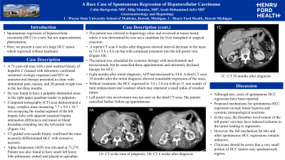
Case Description/Methods: A 73-year-old man with a past medical history of hepatitis C (treated with confirmed sustained virologic response) and HIV infection well controlled on antiretroviral therapy presented to clinic with abdominal pain, nausea and 20-pound weight loss over three months. He was found to have a palpable abdominal mass in the right upper quadrant tender to palpation. Computed tomography (CT) demonstrated a large, complex mass measuring 7.7 x 9.0 x 10.7 cm occupying the medial segment of the left hepatic lobe with a combination of tumor and bland thrombus extending into the left portal vein (Figure 1A). Alpha fetoprotein (AFP) was elevated at 73,279 ng/mL. CT-guided core needle biopsy demonstrated poorly-differentiated HCC with extensive necrosis. Liver synthetic function was preserved with no evidence of cirrhosis on transient elastography. Patient was also found to have small left lower lobe pulmonary emboli and placed on apixaban. The patient was not a candidate for liver transplant or surgical resection. A repeat CT scan 6 weeks after diagnosis showed interval decrease in the mass to 7.6 x 5.8 x 8.6 cm but with continued extension into the left portal vein (Figure 1B). The patient was scheduled for systemic therapy with atezolizumab and bevacizumab, but he declined therapy for HCC. Eight months after initial diagnosis, AFP had decreased to 54.8 ng/dL. A third CT scan 10 months after the initial diagnosis showed remarkable regression of the mass (Figure 1C). Without treatment, the HCC regressed to 3.0 x 2.6 cm with an 11 mm nodule of faint enhancement and washout consistent with a small nidus of residual tumor. Left portal vein involvement was no longer seen. The patient declined further follow-up.
Discussion: Although rare, cases of spontaneous HCC regression have been reported. Proposed mechanisms for spontaneous HCC regression include tumor hypoxia and systemic immunological reactions. In this case, necrosis was seen on the biopsy suggesting tumor regression may have already been occurring at the time of diagnosis potentially from ischemia. The primary blood supply for HCC is the hepatic artery, but the tumor may have outgrown its arterial supply or tumor invasion may have disrupted the artery. Thrombus involvement of the left portal vein may have further contributed to coagulative necrosis.

Disclosures:
Colin Hartgerink, BS1, Dilip Moonka, MD2, Syed-Mohammed Jafri, MD2. P1355 - A Rare Case of Spontaneous Regression of Hepatocellular Carcinoma, ACG 2024 Annual Scientific Meeting Abstracts. Philadelphia, PA: American College of Gastroenterology.
Colin Hartgerink, BS1, Dilip Moonka, MD2, Syed-Mohammed Jafri, MD2
1Wayne State University School of Medicine, Detroit, MI; 2Henry Ford Health, Detroit, MI
Introduction: Spontaneous regression of hepatocellular carcinoma (HCC) is a rare, but not unprecedented, phenomenon. Here, we present a case of a large HCC tumor which regressed without treatment.
Case Description/Methods: A 73-year-old man with a past medical history of hepatitis C (treated with confirmed sustained virologic response) and HIV infection well controlled on antiretroviral therapy presented to clinic with abdominal pain, nausea and 20-pound weight loss over three months. He was found to have a palpable abdominal mass in the right upper quadrant tender to palpation. Computed tomography (CT) demonstrated a large, complex mass measuring 7.7 x 9.0 x 10.7 cm occupying the medial segment of the left hepatic lobe with a combination of tumor and bland thrombus extending into the left portal vein (Figure 1A). Alpha fetoprotein (AFP) was elevated at 73,279 ng/mL. CT-guided core needle biopsy demonstrated poorly-differentiated HCC with extensive necrosis. Liver synthetic function was preserved with no evidence of cirrhosis on transient elastography. Patient was also found to have small left lower lobe pulmonary emboli and placed on apixaban. The patient was not a candidate for liver transplant or surgical resection. A repeat CT scan 6 weeks after diagnosis showed interval decrease in the mass to 7.6 x 5.8 x 8.6 cm but with continued extension into the left portal vein (Figure 1B). The patient was scheduled for systemic therapy with atezolizumab and bevacizumab, but he declined therapy for HCC. Eight months after initial diagnosis, AFP had decreased to 54.8 ng/dL. A third CT scan 10 months after the initial diagnosis showed remarkable regression of the mass (Figure 1C). Without treatment, the HCC regressed to 3.0 x 2.6 cm with an 11 mm nodule of faint enhancement and washout consistent with a small nidus of residual tumor. Left portal vein involvement was no longer seen. The patient declined further follow-up.
Discussion: Although rare, cases of spontaneous HCC regression have been reported. Proposed mechanisms for spontaneous HCC regression include tumor hypoxia and systemic immunological reactions. In this case, necrosis was seen on the biopsy suggesting tumor regression may have already been occurring at the time of diagnosis potentially from ischemia. The primary blood supply for HCC is the hepatic artery, but the tumor may have outgrown its arterial supply or tumor invasion may have disrupted the artery. Thrombus involvement of the left portal vein may have further contributed to coagulative necrosis.

Figure: 1A: CT of the mass at the time of diagnosis; 1B: CT of the mass 6 weeks after diagnosis; 1C: CT of the mass 10 months after diagnosis
Disclosures:
Colin Hartgerink indicated no relevant financial relationships.
Dilip Moonka: AbbVie – Speakers Bureau. Gilead – Speakers Bureau. Intercept – Speakers Bureau. Madrigal – Speakers Bureau.
Syed-Mohammed Jafri: Gilead, Takeda, Abbvie, Intercept, VectivBio – Advisor or Review Panel Member, Speakers Bureau.
Colin Hartgerink, BS1, Dilip Moonka, MD2, Syed-Mohammed Jafri, MD2. P1355 - A Rare Case of Spontaneous Regression of Hepatocellular Carcinoma, ACG 2024 Annual Scientific Meeting Abstracts. Philadelphia, PA: American College of Gastroenterology.


