Sunday Poster Session
Category: Liver
P1228 - Comparing FibroScan at Two Rib Spaces With Liver Histology for Accurate Fibrosis Staging in MASLD Patients
Sunday, October 27, 2024
3:30 PM - 7:00 PM ET
Location: Exhibit Hall E

Has Audio

Jena Velji-Ibrahim, MD, MSc
Prisma Health-Upstate/University of South Carolina School of Medicine
Greenville, SC
Presenting Author(s)
Jena Velji-Ibrahim, MD, MSc1, Jordan Woodard, MD2, Gary Abrams, MD3
1Prisma Health-Upstate/University of South Carolina School of Medicine, Greenville, SC; 2University of Kentucky College of Medicine, Lexington, KY; 3Prisma Health, University of South Carolina School of Medicine, Hoover, AL
Introduction: Metabolic dysfunction-associated steatotic liver disease (MASLD) is increasingly prevalent in the United States. Vibration-controlled transient elastography using FibroScan is a common non-invasive test for MASLD fibrosis. However, the optimal number of intercostal spaces (ICS) for its accuracy, compared to histology has not been determined. We aimed to evaluate FibroScan at different ICSs compared to histology to determine the optimal ICS for accurate fibrosis staging.
Methods: We conducted a retrospective analysis of 181 adults with MASLD who underwent FibroScan at 2 separate ICS and liver biopsy at the same visit. Inclusion criteria included a controlled attenuation parameter (CAP) score of 250+, a minimum of 10 liver stiffness measurements (LSM, kPa) at both rib spaces, IQR/med < 30%, and a liver biopsy with at least 10 portal tracts. FibroScan fibrosis stages were categorized as follows: kPa < 5 (stage 0), 5-7.99 (stage 1) 8-9.99 (stage 2), 10-13.99 (stage 3), and 14+ (stage 4). Histological fibrosis staging was defined using the METAVIR scoring system. LSM had to be within 1 stage of fibrosis on liver biopsy to agree. Statistical analysis was completed via SPSS v28.
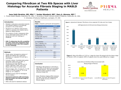
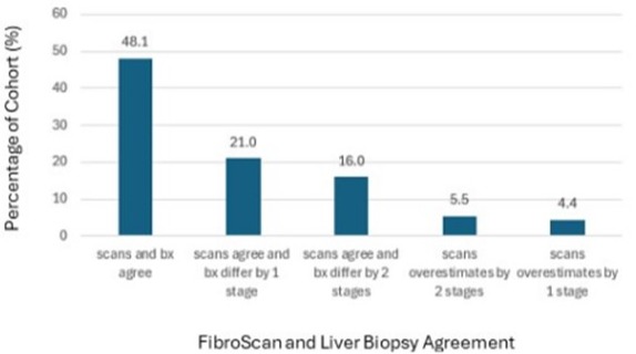
Results: Participants had an average age of 54.3±14 years, with a BMI of 34.6±8 kg/m2, comprising 86.2% white, 7.6% black, and 4.9% Hispanic individuals, with 56.9% females. Among them, 41.6% had T2DM, 57.3% had HTN, and 49.7% had HLD. 71.6% were obese. FibroScan fibrosis stage was stage 0-1 in 48.3%, stage 2 in 10.7%, stage 3 in 15.7%, and stage 4 in 25.2%. As shown in Figure 1, when both ICS sites showed the same fibrosis stage, 48.1% of subjects had the correct stage confirmed on biopsy. In 21.0% and 16.0% of cases, both sites displayed the same stage but differed from biopsy by 1 stage and 2 stages, respectively. FibroScan kPa values at 2 ICS sites differed by 1 stage in 46 (25.4%) and 2 stages in 10 (5.5%) subjects compared to biopsy. Overall, the lower kPa measurements were correct in determining fibrosis within 1 stage in 48/56 (86%). Lower kPa was correct in 38/46 (83%) subjects with 1 stage difference and 10/10 (100%) with 2 stage difference.
Discussion: FibroScan LSM accurately predicted stages of fibrosis at 2 separate ICSs about 50% which is consistent with prior positive predictive values studies utilizing a single ICS. When FibroScan staging is different between ICSs, using the lower kPa measurement has greater accuracy for fibrosis staging.

Disclosures:
Jena Velji-Ibrahim, MD, MSc1, Jordan Woodard, MD2, Gary Abrams, MD3. P1228 - Comparing FibroScan at Two Rib Spaces With Liver Histology for Accurate Fibrosis Staging in MASLD Patients, ACG 2024 Annual Scientific Meeting Abstracts. Philadelphia, PA: American College of Gastroenterology.
1Prisma Health-Upstate/University of South Carolina School of Medicine, Greenville, SC; 2University of Kentucky College of Medicine, Lexington, KY; 3Prisma Health, University of South Carolina School of Medicine, Hoover, AL
Introduction: Metabolic dysfunction-associated steatotic liver disease (MASLD) is increasingly prevalent in the United States. Vibration-controlled transient elastography using FibroScan is a common non-invasive test for MASLD fibrosis. However, the optimal number of intercostal spaces (ICS) for its accuracy, compared to histology has not been determined. We aimed to evaluate FibroScan at different ICSs compared to histology to determine the optimal ICS for accurate fibrosis staging.
Methods: We conducted a retrospective analysis of 181 adults with MASLD who underwent FibroScan at 2 separate ICS and liver biopsy at the same visit. Inclusion criteria included a controlled attenuation parameter (CAP) score of 250+, a minimum of 10 liver stiffness measurements (LSM, kPa) at both rib spaces, IQR/med < 30%, and a liver biopsy with at least 10 portal tracts. FibroScan fibrosis stages were categorized as follows: kPa < 5 (stage 0), 5-7.99 (stage 1) 8-9.99 (stage 2), 10-13.99 (stage 3), and 14+ (stage 4). Histological fibrosis staging was defined using the METAVIR scoring system. LSM had to be within 1 stage of fibrosis on liver biopsy to agree. Statistical analysis was completed via SPSS v28.
Results: Participants had an average age of 54.3±14 years, with a BMI of 34.6±8 kg/m2, comprising 86.2% white, 7.6% black, and 4.9% Hispanic individuals, with 56.9% females. Among them, 41.6% had T2DM, 57.3% had HTN, and 49.7% had HLD. 71.6% were obese. FibroScan fibrosis stage was stage 0-1 in 48.3%, stage 2 in 10.7%, stage 3 in 15.7%, and stage 4 in 25.2%. As shown in Figure 1, when both ICS sites showed the same fibrosis stage, 48.1% of subjects had the correct stage confirmed on biopsy. In 21.0% and 16.0% of cases, both sites displayed the same stage but differed from biopsy by 1 stage and 2 stages, respectively. FibroScan kPa values at 2 ICS sites differed by 1 stage in 46 (25.4%) and 2 stages in 10 (5.5%) subjects compared to biopsy. Overall, the lower kPa measurements were correct in determining fibrosis within 1 stage in 48/56 (86%). Lower kPa was correct in 38/46 (83%) subjects with 1 stage difference and 10/10 (100%) with 2 stage difference.
Discussion: FibroScan LSM accurately predicted stages of fibrosis at 2 separate ICSs about 50% which is consistent with prior positive predictive values studies utilizing a single ICS. When FibroScan staging is different between ICSs, using the lower kPa measurement has greater accuracy for fibrosis staging.

Figure: Figure 1. Agreement between FibroScan at 2 separate intercostal space sites and liver biopsy.
Disclosures:
Jena Velji-Ibrahim indicated no relevant financial relationships.
Jordan Woodard indicated no relevant financial relationships.
Gary Abrams indicated no relevant financial relationships.
Jena Velji-Ibrahim, MD, MSc1, Jordan Woodard, MD2, Gary Abrams, MD3. P1228 - Comparing FibroScan at Two Rib Spaces With Liver Histology for Accurate Fibrosis Staging in MASLD Patients, ACG 2024 Annual Scientific Meeting Abstracts. Philadelphia, PA: American College of Gastroenterology.
