Sunday Poster Session
Category: GI Bleeding
P0798 - A Case of Rectal Endometriosis Manifesting as Cyclic Hematochezia
Sunday, October 27, 2024
3:30 PM - 7:00 PM ET
Location: Exhibit Hall E

Has Audio
- IT
Isra N. Taha, MD
Saint Louis University School of Medicine
St. Louis, MO
Presenting Author(s)
Isra N. Taha, MD1, Mahmoud Y. Madi, MD2, Radhika Patel, MD2, Antonio R. Cheesman, MD2, Danielle Carpenter, MD2
1Saint Louis University School of Medicine, St. Louis, MO; 2SSM Health Saint Louis University Hospital, St. Louis, MO
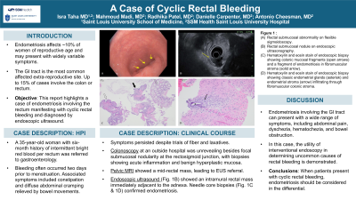
Introduction: Endometriosis, defined as the growth of uterine lining outside the uterus, affects around 10% of women of reproductive age and may cause inflammation, pain, or infertility. While endometriosis most commonly involves reproductive organs and nearby connective tissues, up to 15% of cases may involve the colon or rectum and presentations can vary widely. We present a case of endometriosis involving the rectal submucosa and manifesting as cyclic rectal bleeding in a young female patient.
Case Description/Methods: A 35-year-old woman with six-month history of intermittent bright red blood in her stool was referred to gastroenterology by her primary care physician. She noted rectal bleeding often occurring two days prior to menstruation, and occasional constipation and abdominal cramping relieved by bowel movements. Prior medical history and family history were non-contributory.
Initially, she was treated with a trial of fiber and laxatives. However, her symptoms persisted, prompting further work-up. Colonoscopy revealed a tortuous rectosigmoid junction with an ulcerated area, biopsies of which showed acute inflammation with focal superficial hyperplastic changes.
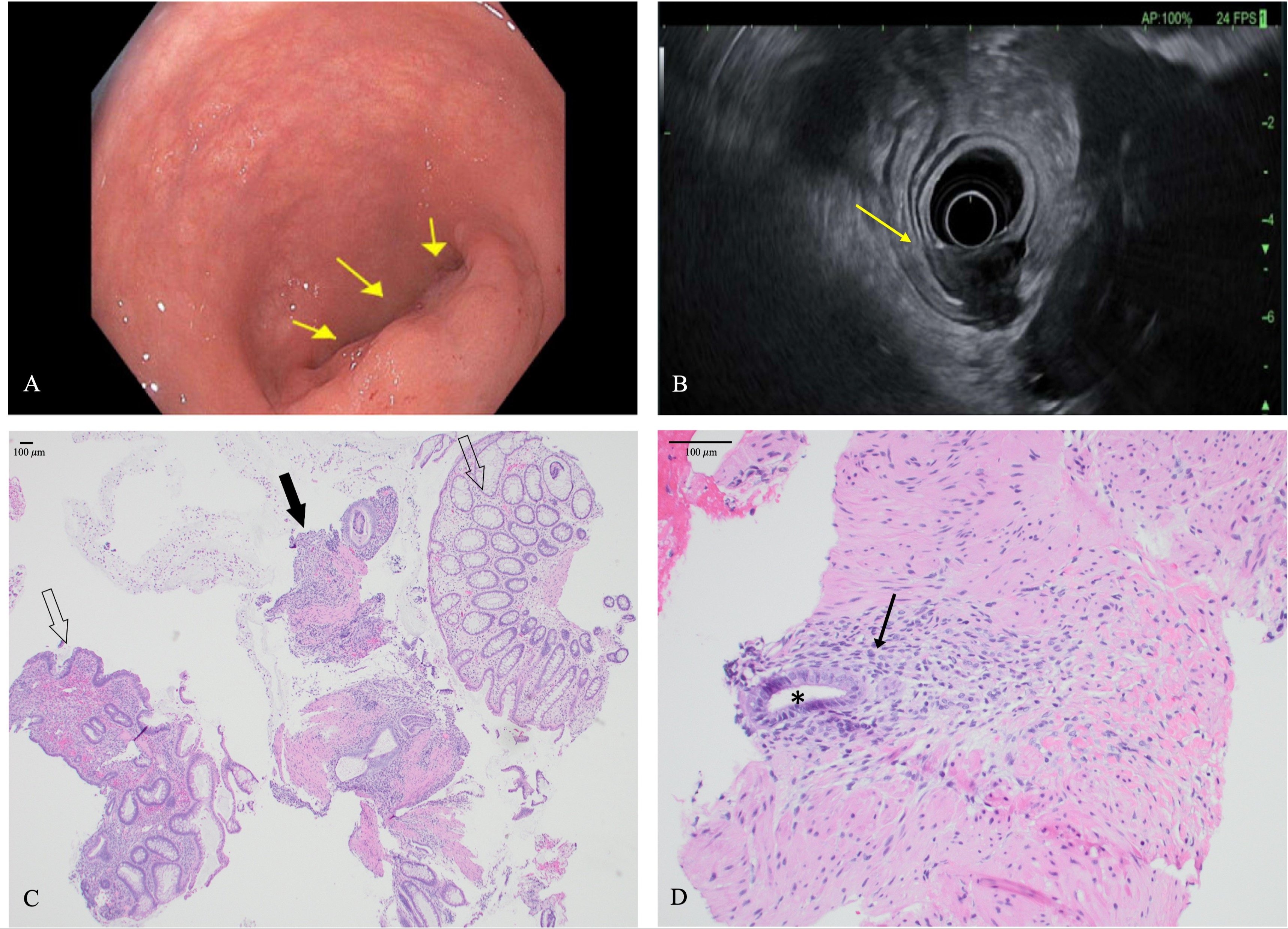
Given the abnormalities on initial colonoscopy and the persistence of rectal bleeding over the next six months, follow-up flexible sigmoidoscopy was completed. This revealed a protruding submucosal nodule (Figure 1A); biopsies showed benign hyperplastic mucosa with absence of submucosa in the specimen. Pelvic MRI was obtained to evaluate for the existence of a submucosal mass; imaging showed a mid-rectal mass and a fibroid-appearing uterus. Further evaluation with endoscopic ultrasound (Figure 1B) and rectal lesion biopsies revealed endometriosis: the pathology highlighted bland endometrial stroma and glands infiltrating between smooth muscle fibers (Figures 1C and 1D). Immunostains confirmed the diagnosis, with positive staining of glands by ER and PAX8 and positive staining of stroma by ER and CD10.
Discussion: In this case of rectal endometriosis, the utility of interventional endoscopy in determining uncommon causes of rectal bleeding is demonstrated. Although the gastrointestinal system is the most commonly affected extra-reproductive site in endometriosis, the presentation can range from asymptomatic to hematochezia to bowel obstruction in severe cases. We see in this case that when patients present with cyclic rectal bleeding, endometriosis should be considered in the differential and abnormal findings should be evaluated further.

Disclosures:
Isra N. Taha, MD1, Mahmoud Y. Madi, MD2, Radhika Patel, MD2, Antonio R. Cheesman, MD2, Danielle Carpenter, MD2. P0798 - A Case of Rectal Endometriosis Manifesting as Cyclic Hematochezia, ACG 2024 Annual Scientific Meeting Abstracts. Philadelphia, PA: American College of Gastroenterology.
1Saint Louis University School of Medicine, St. Louis, MO; 2SSM Health Saint Louis University Hospital, St. Louis, MO
Introduction: Endometriosis, defined as the growth of uterine lining outside the uterus, affects around 10% of women of reproductive age and may cause inflammation, pain, or infertility. While endometriosis most commonly involves reproductive organs and nearby connective tissues, up to 15% of cases may involve the colon or rectum and presentations can vary widely. We present a case of endometriosis involving the rectal submucosa and manifesting as cyclic rectal bleeding in a young female patient.
Case Description/Methods: A 35-year-old woman with six-month history of intermittent bright red blood in her stool was referred to gastroenterology by her primary care physician. She noted rectal bleeding often occurring two days prior to menstruation, and occasional constipation and abdominal cramping relieved by bowel movements. Prior medical history and family history were non-contributory.
Initially, she was treated with a trial of fiber and laxatives. However, her symptoms persisted, prompting further work-up. Colonoscopy revealed a tortuous rectosigmoid junction with an ulcerated area, biopsies of which showed acute inflammation with focal superficial hyperplastic changes.
Given the abnormalities on initial colonoscopy and the persistence of rectal bleeding over the next six months, follow-up flexible sigmoidoscopy was completed. This revealed a protruding submucosal nodule (Figure 1A); biopsies showed benign hyperplastic mucosa with absence of submucosa in the specimen. Pelvic MRI was obtained to evaluate for the existence of a submucosal mass; imaging showed a mid-rectal mass and a fibroid-appearing uterus. Further evaluation with endoscopic ultrasound (Figure 1B) and rectal lesion biopsies revealed endometriosis: the pathology highlighted bland endometrial stroma and glands infiltrating between smooth muscle fibers (Figures 1C and 1D). Immunostains confirmed the diagnosis, with positive staining of glands by ER and PAX8 and positive staining of stroma by ER and CD10.
Discussion: In this case of rectal endometriosis, the utility of interventional endoscopy in determining uncommon causes of rectal bleeding is demonstrated. Although the gastrointestinal system is the most commonly affected extra-reproductive site in endometriosis, the presentation can range from asymptomatic to hematochezia to bowel obstruction in severe cases. We see in this case that when patients present with cyclic rectal bleeding, endometriosis should be considered in the differential and abnormal findings should be evaluated further.

Figure: Figure 1: (A) Rectal submucosal abnormality on flexible sigmoidoscopy. (B) Rectal submucosal nodule on endoscopic ultrasonography. (C) Hematoxylin and eosin stain of endoscopic biopsy showing colonic mucosal fragments (open arrows) and a fragment of endometriosis in fibromuscular stroma (solid arrow). (D) Hematoxylin and eosin stain of endoscopic biopsy showing classic endometrial glands (asterisk) and endometrial stroma (arrow) infiltrating through fibromuscular colonic stroma.
Disclosures:
Isra Taha indicated no relevant financial relationships.
Mahmoud Madi indicated no relevant financial relationships.
Radhika Patel indicated no relevant financial relationships.
Antonio Cheesman indicated no relevant financial relationships.
Danielle Carpenter indicated no relevant financial relationships.
Isra N. Taha, MD1, Mahmoud Y. Madi, MD2, Radhika Patel, MD2, Antonio R. Cheesman, MD2, Danielle Carpenter, MD2. P0798 - A Case of Rectal Endometriosis Manifesting as Cyclic Hematochezia, ACG 2024 Annual Scientific Meeting Abstracts. Philadelphia, PA: American College of Gastroenterology.
