Sunday Poster Session
Category: General Endoscopy
P0711 - Endoscopic Management of Foreign Body Perforating Through Hiatal Hernia Into Thoracic Cavity
Sunday, October 27, 2024
3:30 PM - 7:00 PM ET
Location: Exhibit Hall E

Has Audio

Fathima K. Suhail, BS, MD
SUNY Upstate Medical University
Syracuse, NY
Presenting Author(s)
Award: Presidential Poster Award
Fathima K. Suhail, BS, MD, Kelita Singh, MD
SUNY Upstate Medical University, Syracuse, NY
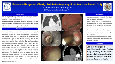
Introduction: Most ingested foreign bodies pass spontaneously,however, up to 20% require endoscopic removal. Less than 1% require surgical intervention when complicated by perforation or fistula formation. We present a case of a foreign body that fistulized from the stomach into the pleural cavity, forming an infected pleural collection that was managed endoscopically.
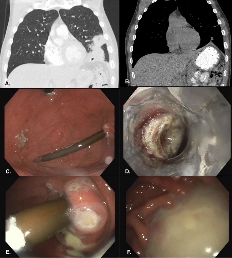
Case Description/Methods: A 44-year-old incarcerated male presented with fevers and left upper quadrant pain for 2 weeks. He swallowed a pen 1 week prior. He was febrile and saturating at 92% on room air, otherwise stable. He appeared comfortable without peritonitic signs. Labs revealed a leukocytosis of 12.9. CT showed a pen in the stomach coursing through the diaphragm, into a left pleural collection and terminating in the fifth intercostal space. A combined case with surgery was planned for endoscopic removal of the pen, closure of the gastro-pleural fistula with surgical drainage of the pleural abscess. We dislodged the pen from the lesser curvature side with a snare. The defect was closed with APC and a padlock over-the-scope clip. We then dislodged the pen from the site of perforation after which copious pus drained through the fistulous tract. The pen was removed from the patient using a snare. After discussion with surgery, we left the gastric side of the perforation open for drainage of the collection into the stomach to facilitate source control. The patient did well post-procedurally. He did not develop a pneumothorax. He continued antibiotics and diet advanced after 48 hours. One month later, CT revealed near resolution of the pleural collection with some oral contrast tracking in the gastric wall. Plan is for elective endoscopic closure.
Discussion: Foreign body ingestions tend to occur in patients with underlying psychiatric diseases, prisoners, and for drug trafficking. 80-90% of foreign bodies pass without intervention. Endoscopy is required in 10-20%. Surgery is required in < 1%. Most foreign bodies in the stomach will pass in 4-6 days. Objects greater than 2-2.5cm in diameter will not pass through the pylorus and longer than 5cm will not pass through the duodenum. Urgent endoscopy is required for sharp objects in the stomach, >5cm in length, and magnets. Complications include perforation and fistula formation which are generally managed surgically. Our case highlights a complication of a large foreign body, fistulizing from a hiatal hernia into the pleural cavity, forming an abscess that was managed endoscopically.

Disclosures:
Fathima K. Suhail, BS, MD, Kelita Singh, MD. P0711 - Endoscopic Management of Foreign Body Perforating Through Hiatal Hernia Into Thoracic Cavity, ACG 2024 Annual Scientific Meeting Abstracts. Philadelphia, PA: American College of Gastroenterology.
Fathima K. Suhail, BS, MD, Kelita Singh, MD
SUNY Upstate Medical University, Syracuse, NY
Introduction: Most ingested foreign bodies pass spontaneously,however, up to 20% require endoscopic removal. Less than 1% require surgical intervention when complicated by perforation or fistula formation. We present a case of a foreign body that fistulized from the stomach into the pleural cavity, forming an infected pleural collection that was managed endoscopically.
Case Description/Methods: A 44-year-old incarcerated male presented with fevers and left upper quadrant pain for 2 weeks. He swallowed a pen 1 week prior. He was febrile and saturating at 92% on room air, otherwise stable. He appeared comfortable without peritonitic signs. Labs revealed a leukocytosis of 12.9. CT showed a pen in the stomach coursing through the diaphragm, into a left pleural collection and terminating in the fifth intercostal space. A combined case with surgery was planned for endoscopic removal of the pen, closure of the gastro-pleural fistula with surgical drainage of the pleural abscess. We dislodged the pen from the lesser curvature side with a snare. The defect was closed with APC and a padlock over-the-scope clip. We then dislodged the pen from the site of perforation after which copious pus drained through the fistulous tract. The pen was removed from the patient using a snare. After discussion with surgery, we left the gastric side of the perforation open for drainage of the collection into the stomach to facilitate source control. The patient did well post-procedurally. He did not develop a pneumothorax. He continued antibiotics and diet advanced after 48 hours. One month later, CT revealed near resolution of the pleural collection with some oral contrast tracking in the gastric wall. Plan is for elective endoscopic closure.
Discussion: Foreign body ingestions tend to occur in patients with underlying psychiatric diseases, prisoners, and for drug trafficking. 80-90% of foreign bodies pass without intervention. Endoscopy is required in 10-20%. Surgery is required in < 1%. Most foreign bodies in the stomach will pass in 4-6 days. Objects greater than 2-2.5cm in diameter will not pass through the pylorus and longer than 5cm will not pass through the duodenum. Urgent endoscopy is required for sharp objects in the stomach, >5cm in length, and magnets. Complications include perforation and fistula formation which are generally managed surgically. Our case highlights a complication of a large foreign body, fistulizing from a hiatal hernia into the pleural cavity, forming an abscess that was managed endoscopically.

Figure: Figure 1: A - CT scan showing foreign body fistulizing from stomach into pleural cavity/collection. B - CT scan 1 month following foreign body removal with near resolution of pleural collection. C - Foreign body (pen) in large hiatal hernia penetrating through gastric wall. D - Closure of lesser curvature side of defect with padlock over-the-scope clip. E - Apparent site of full thickness perforation. F - Pus draining into stomach after removal of pen.
Disclosures:
Fathima Suhail indicated no relevant financial relationships.
Kelita Singh indicated no relevant financial relationships.
Fathima K. Suhail, BS, MD, Kelita Singh, MD. P0711 - Endoscopic Management of Foreign Body Perforating Through Hiatal Hernia Into Thoracic Cavity, ACG 2024 Annual Scientific Meeting Abstracts. Philadelphia, PA: American College of Gastroenterology.


