Sunday Poster Session
Category: General Endoscopy
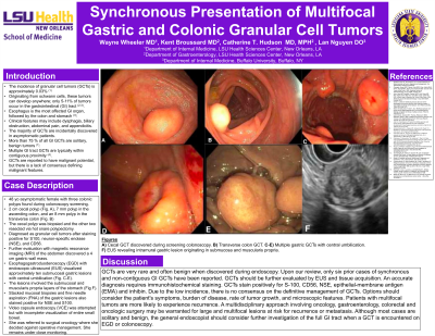
P0719 - Synchronous Presentation of Multifocal Gastric and Colonic Granular Cell Tumors
Sunday, October 27, 2024
3:30 PM - 7:00 PM ET
Location: Exhibit Hall E

Has Audio

Austin Wayne Wayne Wheeler, MD
LSU Health New Orleans School of Medicine
New Orleans, LA
Presenting Author(s)
Austin W. Wheeler, MD1, Kent Broussard, MD2, Catherine T.. Hudson, MD, MPH2
1LSU Health New Orleans School of Medicine, New Orleans, LA; 2LSU, New Orleans, LA
Introduction: The incidence of granular cell tumors (GCTs) is estimated at 0.03%. Originating from schwann cells, GCTs are predominantly found in the oral cavity, subcutaneous tissue, or on the skin. The esophagus is the most affected gastrointestinal (GI) site, followed by the colon and stomach. More than 75 % of all GI GCTs are solitary, benign tumors. GCTs have been reported to have malignant potential, but there is a lack of consensus defining malignant features.
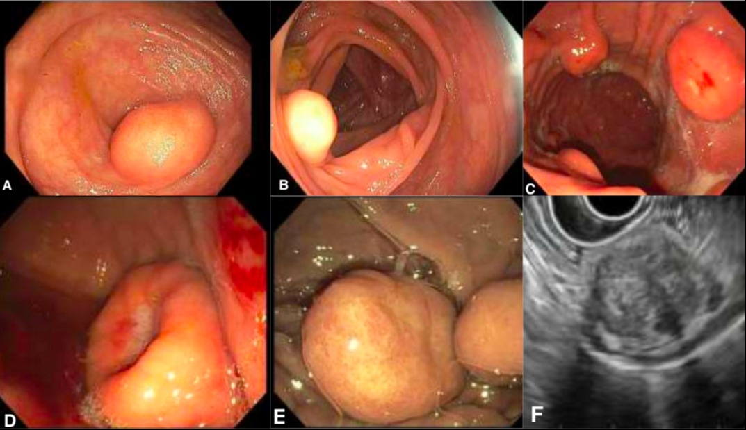
Case Description/Methods: A 48-year-old female was found to have three large colonic polyps during colonoscopy. These included a 2 cm cecal polyp (Fig A), a 7 mm ascending colon polyp, and an 8 mm transverse colon polyp (Fig B). The cecal polyp was biopsied and the other two resected via hot snare. The polyps were identified as GCTs after staining positive for S100, neuron-specific enolase (NSE), and CD56. Further workup with magnetic resonance imaging (MRI) of the abdomen discovered a 4 cm gastric wall mass. Esophagogastroduodenoscopy (EGD) with endoscopic ultrasound (EUS) was performed and visualized approximately ten umbilicated lesions in the stomach involving the submucosal and muscularis propria layers (Figs C-F). Stacked biopsies and fine needle aspiration (FNA) of the gastric lesions again stained positive for GCTs. She is being followed closely by a multidisciplinary team while considering her options for treatment.
Discussion: GCTs are very rare and often benign when discovered during endoscopy. Upon our review, only six prior cases of synchronous and non-contiguous GI GCTs have been reported. GCTs should be further evaluated by EUS and tissue acquisition. An accurate diagnosis requires immunohistochemical staining. GCTs stain positively for S-100, CD56, NSE, epithelial-membrane antigen (EMA) and inhibin. Due to the low incidence, there is no consensus on the definitive management of GCTs. Options should consider the patient's symptoms, burden of disease, rate of tumor growth, and microscopic features. Patients with multifocal tumors are more likely to experience recurrence. A multidisciplinary approach involving oncology, gastroenterology, colorectal and oncologic surgery may be warranted for large and multifocal lesions at risk for recurrence or metastasis. Although most cases are solitary and benign, the general endoscopist should consider further investigation of the full GI tract when a GCT is encountered on EGD or colonoscopy.

Disclosures:
Austin W. Wheeler, MD1, Kent Broussard, MD2, Catherine T.. Hudson, MD, MPH2. P0719 - Synchronous Presentation of Multifocal Gastric and Colonic Granular Cell Tumors, ACG 2024 Annual Scientific Meeting Abstracts. Philadelphia, PA: American College of Gastroenterology.
1LSU Health New Orleans School of Medicine, New Orleans, LA; 2LSU, New Orleans, LA
Introduction: The incidence of granular cell tumors (GCTs) is estimated at 0.03%. Originating from schwann cells, GCTs are predominantly found in the oral cavity, subcutaneous tissue, or on the skin. The esophagus is the most affected gastrointestinal (GI) site, followed by the colon and stomach. More than 75 % of all GI GCTs are solitary, benign tumors. GCTs have been reported to have malignant potential, but there is a lack of consensus defining malignant features.
Case Description/Methods: A 48-year-old female was found to have three large colonic polyps during colonoscopy. These included a 2 cm cecal polyp (Fig A), a 7 mm ascending colon polyp, and an 8 mm transverse colon polyp (Fig B). The cecal polyp was biopsied and the other two resected via hot snare. The polyps were identified as GCTs after staining positive for S100, neuron-specific enolase (NSE), and CD56. Further workup with magnetic resonance imaging (MRI) of the abdomen discovered a 4 cm gastric wall mass. Esophagogastroduodenoscopy (EGD) with endoscopic ultrasound (EUS) was performed and visualized approximately ten umbilicated lesions in the stomach involving the submucosal and muscularis propria layers (Figs C-F). Stacked biopsies and fine needle aspiration (FNA) of the gastric lesions again stained positive for GCTs. She is being followed closely by a multidisciplinary team while considering her options for treatment.
Discussion: GCTs are very rare and often benign when discovered during endoscopy. Upon our review, only six prior cases of synchronous and non-contiguous GI GCTs have been reported. GCTs should be further evaluated by EUS and tissue acquisition. An accurate diagnosis requires immunohistochemical staining. GCTs stain positively for S-100, CD56, NSE, epithelial-membrane antigen (EMA) and inhibin. Due to the low incidence, there is no consensus on the definitive management of GCTs. Options should consider the patient's symptoms, burden of disease, rate of tumor growth, and microscopic features. Patients with multifocal tumors are more likely to experience recurrence. A multidisciplinary approach involving oncology, gastroenterology, colorectal and oncologic surgery may be warranted for large and multifocal lesions at risk for recurrence or metastasis. Although most cases are solitary and benign, the general endoscopist should consider further investigation of the full GI tract when a GCT is encountered on EGD or colonoscopy.

Figure: Figure 1: (A) Cecal GCT. (B) Transverse colon GCT. (C) Multiple GCTs in stomach. (D) Large umbilicated gastric GCT. (E) Pedunculated gastric GCT. (F) Gastric GCT involving submucosal and muscularis propria on EUS.
Disclosures:
Austin Wheeler indicated no relevant financial relationships.
Kent Broussard indicated no relevant financial relationships.
Catherine Hudson indicated no relevant financial relationships.
Austin W. Wheeler, MD1, Kent Broussard, MD2, Catherine T.. Hudson, MD, MPH2. P0719 - Synchronous Presentation of Multifocal Gastric and Colonic Granular Cell Tumors, ACG 2024 Annual Scientific Meeting Abstracts. Philadelphia, PA: American College of Gastroenterology.
