Sunday Poster Session
Category: General Endoscopy
P0721 - Incidental Diagnosis of Metastatic Cervical Cancer via Colonoscopy
Sunday, October 27, 2024
3:30 PM - 7:00 PM ET
Location: Exhibit Hall E

Has Audio
- DW
Daniel Weinblatt, DO
Reading Hospital - Tower Health
West Reading, PA
Presenting Author(s)
Daniel Weinblatt, DO1, Adam Spiegel, DO2
1Reading Hospital - Tower Health, West Reading, PA; 2US Digestive Health, West Reading, PA
Introduction: Iron deficiency anemia is a common indication for bidirectional endoscopy. The following case report describes the incidental discovery of metastatic cervical cancer during a colonoscopy that was performed for iron deficiency anemia.
Case Description/Methods: An 81 year old woman with a history of restless leg syndrome presented to the hospital due to iron deficiency anemia discovered on outpatient labs. She had labs ordered due to fatigue which revealed Hgb 6.5 g/dL with MCV 80.4 fL, as well as ferritin 3 ng/mL, and iron saturation 2%. A previous Hgb was 13.8 g/dL in 2016. She reported fatigue but otherwise a review of systems was negative. Her most recent colonoscopy five years prior had revealed moderate pancolonic diverticulosis and grade I internal hemorrhoids. She had never had an upper endoscopy. Vital signs were within normal limits and physical examination was unremarkable.
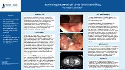
She underwent EGD and colonoscopy the following day. The EGD was normal. On digital rectal exam prior to colonoscopy there was a firm, nodular mass-like lesion palpated along the anterior and left lateral wall of the rectum. On colonoscopy, there was a 2 cm friable and firm mass like area in the distal rectum just proximal to the dentate line. There was no intraluminal mass in this location, which raised concern for extrinsic invasion. The area was biopsied in a tunneled fashion. The remainder of the colonoscopy revealed diverticulosis and internal hemorrhoids. Subsequent discussion with the patient and her daughter revealed that the patient had been experiencing vaginal bleeding since two months prior to the admission. Rectal biopsies revealed squamous cell carcinoma. The patient underwent CT which demonstrated an abnormal masslike thickening of the upper vaginal vault and cervix, with tissue contacting the rectum, concerning for gynecologic malignancy. She then underwent a pelvic exam which revealed a cervical mass. Biopsies confirmed invasive squamous cell carcinoma. She is now planned for chemotherapy and radiation for stage IVb cervical cancer.
Discussion: This case describes an unexpected diagnosis of metastatic cervical cancer during a colonoscopy that was performed for iron deficiency anemia. The case highlights the importance of conducting a thorough history and physical, and the need to investigate gynecologic sources of iron deficiency when appropriate.

Disclosures:
Daniel Weinblatt, DO1, Adam Spiegel, DO2. P0721 - Incidental Diagnosis of Metastatic Cervical Cancer via Colonoscopy, ACG 2024 Annual Scientific Meeting Abstracts. Philadelphia, PA: American College of Gastroenterology.
1Reading Hospital - Tower Health, West Reading, PA; 2US Digestive Health, West Reading, PA
Introduction: Iron deficiency anemia is a common indication for bidirectional endoscopy. The following case report describes the incidental discovery of metastatic cervical cancer during a colonoscopy that was performed for iron deficiency anemia.
Case Description/Methods: An 81 year old woman with a history of restless leg syndrome presented to the hospital due to iron deficiency anemia discovered on outpatient labs. She had labs ordered due to fatigue which revealed Hgb 6.5 g/dL with MCV 80.4 fL, as well as ferritin 3 ng/mL, and iron saturation 2%. A previous Hgb was 13.8 g/dL in 2016. She reported fatigue but otherwise a review of systems was negative. Her most recent colonoscopy five years prior had revealed moderate pancolonic diverticulosis and grade I internal hemorrhoids. She had never had an upper endoscopy. Vital signs were within normal limits and physical examination was unremarkable.
She underwent EGD and colonoscopy the following day. The EGD was normal. On digital rectal exam prior to colonoscopy there was a firm, nodular mass-like lesion palpated along the anterior and left lateral wall of the rectum. On colonoscopy, there was a 2 cm friable and firm mass like area in the distal rectum just proximal to the dentate line. There was no intraluminal mass in this location, which raised concern for extrinsic invasion. The area was biopsied in a tunneled fashion. The remainder of the colonoscopy revealed diverticulosis and internal hemorrhoids. Subsequent discussion with the patient and her daughter revealed that the patient had been experiencing vaginal bleeding since two months prior to the admission. Rectal biopsies revealed squamous cell carcinoma. The patient underwent CT which demonstrated an abnormal masslike thickening of the upper vaginal vault and cervix, with tissue contacting the rectum, concerning for gynecologic malignancy. She then underwent a pelvic exam which revealed a cervical mass. Biopsies confirmed invasive squamous cell carcinoma. She is now planned for chemotherapy and radiation for stage IVb cervical cancer.
Discussion: This case describes an unexpected diagnosis of metastatic cervical cancer during a colonoscopy that was performed for iron deficiency anemia. The case highlights the importance of conducting a thorough history and physical, and the need to investigate gynecologic sources of iron deficiency when appropriate.

Figure: A) Nodular and friable rectal wall tissue noted on colonoscopy. B) Nodular, friable rectal wall tissue seen on retroflexion. C) Irregular masslike thickening of the vagina/cervix contacting the rectum on CT scan.
Disclosures:
Daniel Weinblatt indicated no relevant financial relationships.
Adam Spiegel indicated no relevant financial relationships.
Daniel Weinblatt, DO1, Adam Spiegel, DO2. P0721 - Incidental Diagnosis of Metastatic Cervical Cancer via Colonoscopy, ACG 2024 Annual Scientific Meeting Abstracts. Philadelphia, PA: American College of Gastroenterology.
