Sunday Poster Session
Category: Esophagus
P0589 - Multiple Esophageal Duplication Cysts: A Rare Case of Chronic Dysphagia in Adulthood
Sunday, October 27, 2024
3:30 PM - 7:00 PM ET
Location: Exhibit Hall E

Has Audio
.jpg)
Abdullah Mahmood, MD
University Hospitals Cleveland Medical Center, Case Western Reserve University
Cleveland, OH
Presenting Author(s)
Abdullah Mahmood, MD1, Dayyan Adoor, MD1, Andrew Catanzaro, MD2, Brooke Glessing, MD2
1University Hospitals Cleveland Medical Center, Case Western Reserve University, Cleveland, OH; 2Digestive Health Institute, University Hospitals Cleveland Medical Center, Cleveland, OH
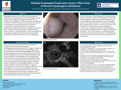
Introduction: Esophageal duplication cysts (EDCs) are rare congenital malformations accounting up to 2.5% of esophageal masses. A large portion of EDCs go undiagnosed due to a lack of symptoms. Presentation in adults is rare as 80% are diagnosed in children. The majority of EDCs arise in the middle and lower third of the esophagus and present with symptoms related to compression of surrounding structures, commonly manifesting as dysphagia, chest pain or epigastric discomfort. EDCs usually occur as single lesions and endoscopically appear similar to a submucosal lesion, with normal appearing esophageal mucosa. We report a unique case of a patient developing multiple EDCs resulting in chronic dysphagia.
Case Description/Methods: A 65 year old female presented with chronic dysphagia exclusive to solid food. The patient had an esophagogastroduodenoscopy (EGD) for epigastric abdominal pain 2 years prior that showed a normal esophagus. Chest Tomography (CT) revealed distal esophageal wall thickening. Barium esophagram was normal. EGD demonstrated multiple longitudinal submucosal nodules extending through the entire length of the esophagus with normal appearing overlying mucosa. Endoscopic ultrasound (EUS) identified 5 round, anechoic, and homogeneous submucosal cysts with well-defined margins that were doppler negative. Mucosal biopsies revealed normal esophageal epithelium. Thoracic surgery evaluation was obtained but given her age and comorbidities, she was to not be a surgical candidate. She was discharged home on a soft diet. Esophageal manometry performed as an outpatient demonstrated normal peristalsis with a single hypercontractile swallow. Impedance study showed complete bolus transit in 80% of swallows. She noted her dysphagia mildly improved on a soft diet.
Discussion: EDCs are thought to arise during gestation between weeks 4-8 due to abnormal budding of the esophagus. EUS provides the best diagnostic utility as it assesses echogenicity and surrounding layers, differentiating from other esophageal masses. Fine needle aspiration can assist as well, however, this has been shown to lead to a 14% rate of infection and is therefore only recommended in cases of diagnostic uncertainty. Management of EDCs is typically surgical, however endoscopic approaches for smaller cysts have been described. Our case is unique as our patient presented during adulthood and was found to have multiple EDCs that developed to become appreciated endoscopically within a 2 year interval.

Note: The table for this abstract can be viewed in the ePoster Gallery section of the ACG 2024 ePoster Site or in The American Journal of Gastroenterology's abstract supplement issue, both of which will be available starting October 27, 2024.
Disclosures:
Abdullah Mahmood, MD1, Dayyan Adoor, MD1, Andrew Catanzaro, MD2, Brooke Glessing, MD2. P0589 - Multiple Esophageal Duplication Cysts: A Rare Case of Chronic Dysphagia in Adulthood, ACG 2024 Annual Scientific Meeting Abstracts. Philadelphia, PA: American College of Gastroenterology.
1University Hospitals Cleveland Medical Center, Case Western Reserve University, Cleveland, OH; 2Digestive Health Institute, University Hospitals Cleveland Medical Center, Cleveland, OH
Introduction: Esophageal duplication cysts (EDCs) are rare congenital malformations accounting up to 2.5% of esophageal masses. A large portion of EDCs go undiagnosed due to a lack of symptoms. Presentation in adults is rare as 80% are diagnosed in children. The majority of EDCs arise in the middle and lower third of the esophagus and present with symptoms related to compression of surrounding structures, commonly manifesting as dysphagia, chest pain or epigastric discomfort. EDCs usually occur as single lesions and endoscopically appear similar to a submucosal lesion, with normal appearing esophageal mucosa. We report a unique case of a patient developing multiple EDCs resulting in chronic dysphagia.
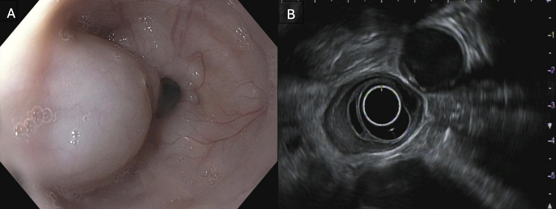
Case Description/Methods: A 65 year old female presented with chronic dysphagia exclusive to solid food. The patient had an esophagogastroduodenoscopy (EGD) for epigastric abdominal pain 2 years prior that showed a normal esophagus. Chest Tomography (CT) revealed distal esophageal wall thickening. Barium esophagram was normal. EGD demonstrated multiple longitudinal submucosal nodules extending through the entire length of the esophagus with normal appearing overlying mucosa. Endoscopic ultrasound (EUS) identified 5 round, anechoic, and homogeneous submucosal cysts with well-defined margins that were doppler negative. Mucosal biopsies revealed normal esophageal epithelium. Thoracic surgery evaluation was obtained but given her age and comorbidities, she was to not be a surgical candidate. She was discharged home on a soft diet. Esophageal manometry performed as an outpatient demonstrated normal peristalsis with a single hypercontractile swallow. Impedance study showed complete bolus transit in 80% of swallows. She noted her dysphagia mildly improved on a soft diet.
Discussion: EDCs are thought to arise during gestation between weeks 4-8 due to abnormal budding of the esophagus. EUS provides the best diagnostic utility as it assesses echogenicity and surrounding layers, differentiating from other esophageal masses. Fine needle aspiration can assist as well, however, this has been shown to lead to a 14% rate of infection and is therefore only recommended in cases of diagnostic uncertainty. Management of EDCs is typically surgical, however endoscopic approaches for smaller cysts have been described. Our case is unique as our patient presented during adulthood and was found to have multiple EDCs that developed to become appreciated endoscopically within a 2 year interval.

Figure: A: Esophageal duplication cyst on esophagogastroduodenoscopy (EGD)
B: Esophageal duplication cyst on endoscopic ultrasound (EUS)
B: Esophageal duplication cyst on endoscopic ultrasound (EUS)
Note: The table for this abstract can be viewed in the ePoster Gallery section of the ACG 2024 ePoster Site or in The American Journal of Gastroenterology's abstract supplement issue, both of which will be available starting October 27, 2024.
Disclosures:
Abdullah Mahmood indicated no relevant financial relationships.
Dayyan Adoor indicated no relevant financial relationships.
Andrew Catanzaro indicated no relevant financial relationships.
Brooke Glessing indicated no relevant financial relationships.
Abdullah Mahmood, MD1, Dayyan Adoor, MD1, Andrew Catanzaro, MD2, Brooke Glessing, MD2. P0589 - Multiple Esophageal Duplication Cysts: A Rare Case of Chronic Dysphagia in Adulthood, ACG 2024 Annual Scientific Meeting Abstracts. Philadelphia, PA: American College of Gastroenterology.
