Sunday Poster Session
Category: Esophagus
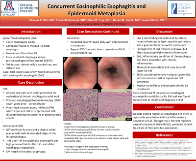
P0591 - Concurrent Eosinophilic Esophagitis and Epidermoid Metaplasia
Sunday, October 27, 2024
3:30 PM - 7:00 PM ET
Location: Exhibit Hall E

Has Audio

Vanessa F. Eller, MD
University of Arizona College of Medicine
Phoenix, AZ
Presenting Author(s)
Vanessa F. Eller, MD1, Elizabeth Rowland, MD1, Brian M.. Fung, MD2, Daniel M.. Jondle, MD3, Joseph David, MD1
1University of Arizona College of Medicine, Phoenix, AZ; 2Arizona Digestive Health, Mesa, AZ; 3GI Alliance, Scottsdale, AZ
Introduction: Epidermoid metaplasia (EM) is a rare esophageal lesion most commonly found in the mid- to distal esophagus, with a prevalence of less than 1%. It is often an incidental finding, but can sometimes be associated with dysphagia and/or gastroesophageal reflux disease (GERD). Known risk factors for this condition include chronic reflux, alcohol use, and tobacco use. In the following case, we present the first case of EM found concurrently with eosinophilic esophagitis (EoE).
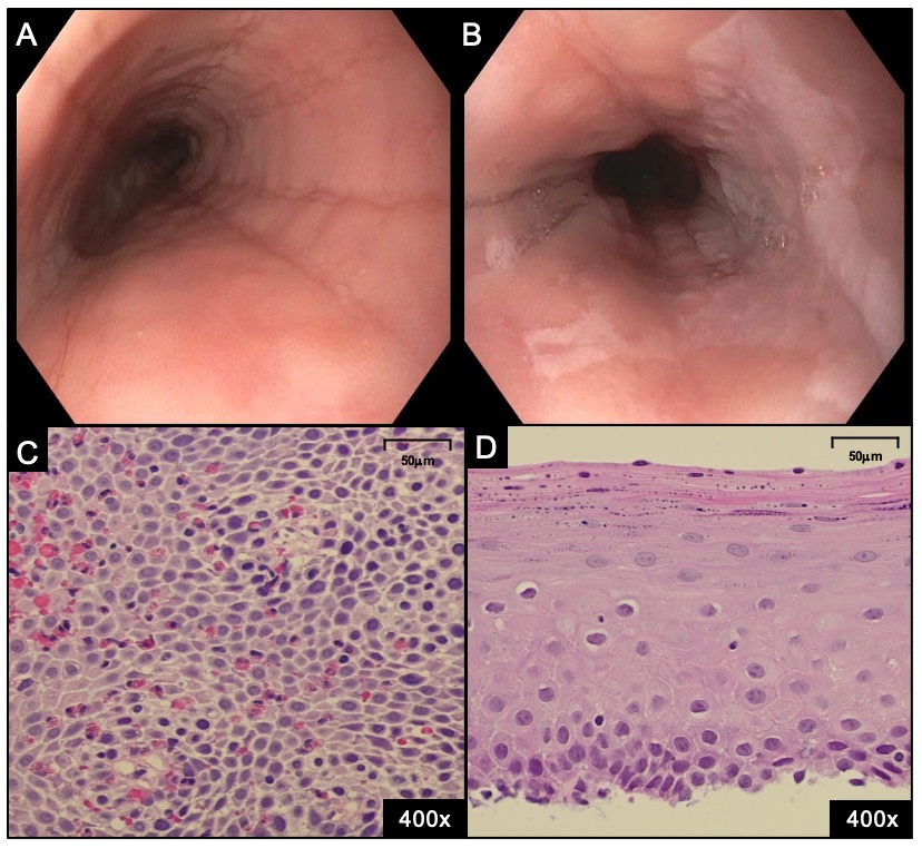
Case Description/Methods: A 63-year-old male with GERD presented for evaluation of chronic dysphagia to solid food. An esophagogastroduodenoscopy (EGD) seven years prior was reportedly unremarkable. The patient was prescribed a proton pump inhibitor (PPI) which improved his reflux symptoms. However, the patient subsequently self-discontinued the PPI due to concerns of potential long-term side effects. A subsequent EGD revealed diffuse linear furrows and a distinct white plaque with well-demarcated edges in the distal esophagus (Figures A & B). Histopathology revealed > 65 and > 40 intraepithelial eosinophils per high-powered field in the mid- and distal esophagus, respectively (Figure C). Additionally, EM was noted in the distal esophagus (Figure D). The patient was restarted on a PPI twice daily with improvement in symptoms. Repeat EGD two months later demonstrated resolution of EoE, but persistent EM.
Discussion: EM is a rare finding characterized by a thick, hyperorthokeratotic layer atop the epithelium and a granular layer below the epithelium. While the pathogenesis of EM remains unknown, chronic inflammation is suspected to play a role in its development. Our patient was found to have EoE at the time of diagnosis of EM. Given that EoE is an inflammatory condition of the esophagus and EM is associated with chronic inflammation, it is theoretically possible that EoE may be a risk factor for EM. More likely, our patient may have had GERD with PPI-responsive esophageal eosinophilia as his risk factor for EM. Despite limited reports of esophageal EM, the condition is considered to have malignant potential, with an increased risk of squamous cell carcinoma. As such, regular surveillance endoscopies should be considered.

Disclosures:
Vanessa F. Eller, MD1, Elizabeth Rowland, MD1, Brian M.. Fung, MD2, Daniel M.. Jondle, MD3, Joseph David, MD1. P0591 - Concurrent Eosinophilic Esophagitis and Epidermoid Metaplasia, ACG 2024 Annual Scientific Meeting Abstracts. Philadelphia, PA: American College of Gastroenterology.
1University of Arizona College of Medicine, Phoenix, AZ; 2Arizona Digestive Health, Mesa, AZ; 3GI Alliance, Scottsdale, AZ
Introduction: Epidermoid metaplasia (EM) is a rare esophageal lesion most commonly found in the mid- to distal esophagus, with a prevalence of less than 1%. It is often an incidental finding, but can sometimes be associated with dysphagia and/or gastroesophageal reflux disease (GERD). Known risk factors for this condition include chronic reflux, alcohol use, and tobacco use. In the following case, we present the first case of EM found concurrently with eosinophilic esophagitis (EoE).
Case Description/Methods: A 63-year-old male with GERD presented for evaluation of chronic dysphagia to solid food. An esophagogastroduodenoscopy (EGD) seven years prior was reportedly unremarkable. The patient was prescribed a proton pump inhibitor (PPI) which improved his reflux symptoms. However, the patient subsequently self-discontinued the PPI due to concerns of potential long-term side effects. A subsequent EGD revealed diffuse linear furrows and a distinct white plaque with well-demarcated edges in the distal esophagus (Figures A & B). Histopathology revealed > 65 and > 40 intraepithelial eosinophils per high-powered field in the mid- and distal esophagus, respectively (Figure C). Additionally, EM was noted in the distal esophagus (Figure D). The patient was restarted on a PPI twice daily with improvement in symptoms. Repeat EGD two months later demonstrated resolution of EoE, but persistent EM.
Discussion: EM is a rare finding characterized by a thick, hyperorthokeratotic layer atop the epithelium and a granular layer below the epithelium. While the pathogenesis of EM remains unknown, chronic inflammation is suspected to play a role in its development. Our patient was found to have EoE at the time of diagnosis of EM. Given that EoE is an inflammatory condition of the esophagus and EM is associated with chronic inflammation, it is theoretically possible that EoE may be a risk factor for EM. More likely, our patient may have had GERD with PPI-responsive esophageal eosinophilia as his risk factor for EM. Despite limited reports of esophageal EM, the condition is considered to have malignant potential, with an increased risk of squamous cell carcinoma. As such, regular surveillance endoscopies should be considered.

Figure: FIGURE: Images from esophagogastroduodenoscopy showing the (A) mid-esophagus with linear furrows consistent with eosinophilic esophagitis (EoE) and (B) the distal esophagus with EoE and concurrent epidermoid metaplasia (EM) as evidenced by the distinct white plaque with well-demarcated edges. Histological images of the esophagus showing (C) EoE changes with intraepithelial eosinophils and (D) EM in the distal esophagus.
Disclosures:
Vanessa Eller indicated no relevant financial relationships.
Elizabeth Rowland indicated no relevant financial relationships.
Brian Fung indicated no relevant financial relationships.
Daniel Jondle indicated no relevant financial relationships.
Joseph David indicated no relevant financial relationships.
Vanessa F. Eller, MD1, Elizabeth Rowland, MD1, Brian M.. Fung, MD2, Daniel M.. Jondle, MD3, Joseph David, MD1. P0591 - Concurrent Eosinophilic Esophagitis and Epidermoid Metaplasia, ACG 2024 Annual Scientific Meeting Abstracts. Philadelphia, PA: American College of Gastroenterology.
