Sunday Poster Session
Category: Esophagus
P0535 - Esophageal Yolk Sac Adenocarcinoma Found Incidentally on Endoscopic Submucosal Dissection: A Rare Coexistence
Sunday, October 27, 2024
3:30 PM - 7:00 PM ET
Location: Exhibit Hall E

Has Audio

Cristina Chiodi, MD
University of Louisville School of Medicine
Sarasota, FL
Presenting Author(s)
Award: Presidential Poster Award
Cristina Chiodi, MD1, David Liu, MD2, Mikayla Hennen, DO2, Dibson Gondhim, MD3, Nathan Liu, MD3
1University of Louisville School of Medicine, Sarasota, FL; 2University of Louisville School of Medicine, Louisville, KY; 3University of Louisville, Louisville, KY
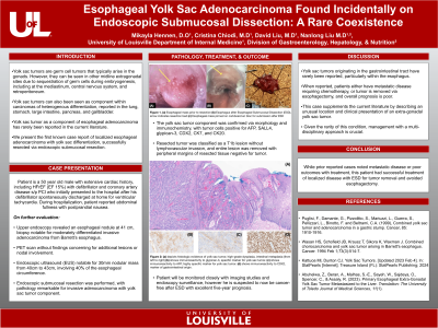
Introduction: Yolk sac tumors (YST) are germ cell tumors that typically arise in the gonads. However, they can be seen in midline extra-gonadal sites due to the sequestration of germ cells during embryogenesis, including mediastinum, central nervous system, and retroperitoneum. It has also been reported as a component within carcinomas of heterogeneous differentiation in the lung, stomach, large intestine, pancreas, and gallbladder. YST as a component of esophageal adenocarcinoma has rarely been reported in current literature. When reported, disease is metastatic requiring chemotherapy, or tumor is removed via esophagectomy. We present the first known case report of localized esophageal adenocarcinoma with yolk sac differentiation, successfully resected via endoscopic submucosal dissection (ESD).
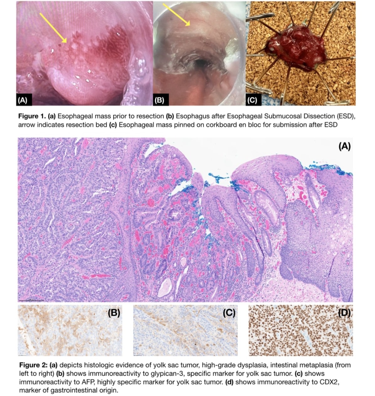
Case Description/Methods: 50-year-old male with an extensive cardiac history, including HFrEF (< 15%) and CAD s/p PCI, who initially presented after defibrillator spontaneously discharged at home for ventricular tachycardia. During hospitalization, he reported abdominal fullness with postprandial nausea. On further evaluation, EGD revealed a nodule at 41 cm with biopsy notable for moderately differentiated invasive adenocarcinoma from Barrett's esophagus. PET scan was negative for additional lesions or nodal involvement. Endoscopic ultrasound (EUS) for further evaluation was performed in the operating room due to his cardiac function, revealing a 30mm nodular mass from 40cm to 43cm involving 40% of the esophageal circumference. During procedure, ESD was performed using a tunnel approach, with pathology confirming adenocarcinoma with YST component with submucosal invasion. The YST component was confirmed via morphology and immunochemistry, with tumor cells positive for AFP, SALL4, glypican-3, CDX2, CK7, and CK20. Resected tumor was classified as T1bN0M0 lesion without lymphovascular invasion. Negative deep and peripheral margins confirmed en-bloc resection- depth of invasion < 500µ. After discussion with surgical and medical oncology, there are plans for endoscopic monitoring and ablation of remaining Barrett's esophagus after cardiac optimization.
Discussion: YSTs originating in the gastrointestinal tract are rarely reported. This case supplements current literature by describing an unusual location and clinical presentation of an extra-gonadal YST. While prior reported cases note metastatic disease or poor outcomes with treatment, this patient had successful treatment of localized disease with ESD for tumor removal.

Disclosures:
Cristina Chiodi, MD1, David Liu, MD2, Mikayla Hennen, DO2, Dibson Gondhim, MD3, Nathan Liu, MD3. P0535 - Esophageal Yolk Sac Adenocarcinoma Found Incidentally on Endoscopic Submucosal Dissection: A Rare Coexistence, ACG 2024 Annual Scientific Meeting Abstracts. Philadelphia, PA: American College of Gastroenterology.
Cristina Chiodi, MD1, David Liu, MD2, Mikayla Hennen, DO2, Dibson Gondhim, MD3, Nathan Liu, MD3
1University of Louisville School of Medicine, Sarasota, FL; 2University of Louisville School of Medicine, Louisville, KY; 3University of Louisville, Louisville, KY
Introduction: Yolk sac tumors (YST) are germ cell tumors that typically arise in the gonads. However, they can be seen in midline extra-gonadal sites due to the sequestration of germ cells during embryogenesis, including mediastinum, central nervous system, and retroperitoneum. It has also been reported as a component within carcinomas of heterogeneous differentiation in the lung, stomach, large intestine, pancreas, and gallbladder. YST as a component of esophageal adenocarcinoma has rarely been reported in current literature. When reported, disease is metastatic requiring chemotherapy, or tumor is removed via esophagectomy. We present the first known case report of localized esophageal adenocarcinoma with yolk sac differentiation, successfully resected via endoscopic submucosal dissection (ESD).
Case Description/Methods: 50-year-old male with an extensive cardiac history, including HFrEF (< 15%) and CAD s/p PCI, who initially presented after defibrillator spontaneously discharged at home for ventricular tachycardia. During hospitalization, he reported abdominal fullness with postprandial nausea. On further evaluation, EGD revealed a nodule at 41 cm with biopsy notable for moderately differentiated invasive adenocarcinoma from Barrett's esophagus. PET scan was negative for additional lesions or nodal involvement. Endoscopic ultrasound (EUS) for further evaluation was performed in the operating room due to his cardiac function, revealing a 30mm nodular mass from 40cm to 43cm involving 40% of the esophageal circumference. During procedure, ESD was performed using a tunnel approach, with pathology confirming adenocarcinoma with YST component with submucosal invasion. The YST component was confirmed via morphology and immunochemistry, with tumor cells positive for AFP, SALL4, glypican-3, CDX2, CK7, and CK20. Resected tumor was classified as T1bN0M0 lesion without lymphovascular invasion. Negative deep and peripheral margins confirmed en-bloc resection- depth of invasion < 500µ. After discussion with surgical and medical oncology, there are plans for endoscopic monitoring and ablation of remaining Barrett's esophagus after cardiac optimization.
Discussion: YSTs originating in the gastrointestinal tract are rarely reported. This case supplements current literature by describing an unusual location and clinical presentation of an extra-gonadal YST. While prior reported cases note metastatic disease or poor outcomes with treatment, this patient had successful treatment of localized disease with ESD for tumor removal.

Figure: Figure 1 (a) shows esophageal mass prior to resection (b) esophagus after resection (c) mass after resection; Figure 2 (a)-(d) show histologic evidence of yolk sac tumor
Disclosures:
Cristina Chiodi indicated no relevant financial relationships.
David Liu indicated no relevant financial relationships.
Mikayla Hennen indicated no relevant financial relationships.
Dibson Gondhim indicated no relevant financial relationships.
Nathan Liu indicated no relevant financial relationships.
Cristina Chiodi, MD1, David Liu, MD2, Mikayla Hennen, DO2, Dibson Gondhim, MD3, Nathan Liu, MD3. P0535 - Esophageal Yolk Sac Adenocarcinoma Found Incidentally on Endoscopic Submucosal Dissection: A Rare Coexistence, ACG 2024 Annual Scientific Meeting Abstracts. Philadelphia, PA: American College of Gastroenterology.


