Sunday Poster Session
Category: Colon
P0367 - Lymphovascular Malformation in the Rectosigmoid Colon
Sunday, October 27, 2024
3:30 PM - 7:00 PM ET
Location: Exhibit Hall E

Has Audio
- GC
Grant Cammock, BS
NYU Grossman School of Medicine
New York, NY
Presenting Author(s)
Grant Cammock, BS1, Gregory Riley, MD1, Renee Williams, MD2
1NYU Grossman School of Medicine, New York, NY; 2NYU Grossman School of Medicine, North Bergen, NJ
Introduction: Lymphovenous malformations (LVM) are one subset of vascular malformations. They arise through abnormal development or acquired defects of lymphatic and venous channels but are rarely in the gastrointestinal tract. This case report presents the findings of an LVM found incidentally in the rectosigmoid colon.
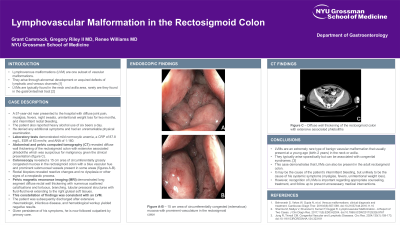
Case Description/Methods: A 57-year-old man presented to the hospital with diffuse joint pain, myalgias, fevers, night sweats, unintentional weight loss for two months, and intermittent rectal bleeding. The patient also reported heavy alcohol use of six beers a day. He denied any additional symptoms and had an unremarkable physical examination. Laboratory tests demonstrated mild normocytic anemia, a CRP of 87.8 mg/L, ESR of 63 mm/hr, and ANA of 1:160. Abdominal and pelvic computed tomography (CT) revealed diffuse wall thickening of the rectosigmoid colon with extensive associated phleboliths which was suspicious for malignancy given the clinical presentation (figure 1, panel A). A colonoscopy revealed a 15 cm area of circumferentially grossly congested mucosa in the rectosigmoid colon with a blue vascular hue and prominent submucosal vessels present in some areas (figure 1, panel B-C). Rectal biopsies revealed reactive changes and no dysplasia or other signs of a neoplastic process. A pelvic magnetic resonance imaging (MRI) demonstrated long segment diffuse rectal wall thickening with numerous scattered calcifications and tortuous, branching, tubular presacral structures with fluid-fluid level extending to the right gluteal soft tissues. This constellation of findings was consistent with an LVM. The patient was subsequently discharged after extensive rheumatologic, infectious disease, and hematological workup yielded negative results. Given persistence of his symptoms, he is now followed outpatient by primary care.
Discussion: LVMs are an extremely rare type of benign vascular malformation that usually present at a young age (birth-2 years) in the neck or axilla. They typically arise sporadically but can be associated with congenital syndromes. This case demonstrates that LVMs can also be present in the adult rectosigmoid colon. It may be the cause of the patient’s intermittent bleeding, but unlikely to be the cause of his systemic symptoms (myalgias, fevers). However, recognition of LVMs is important regarding appropriate counseling, treatment, and follow up to prevent unnecessary medical interventions.

Disclosures:
Grant Cammock, BS1, Gregory Riley, MD1, Renee Williams, MD2. P0367 - Lymphovascular Malformation in the Rectosigmoid Colon, ACG 2024 Annual Scientific Meeting Abstracts. Philadelphia, PA: American College of Gastroenterology.
1NYU Grossman School of Medicine, New York, NY; 2NYU Grossman School of Medicine, North Bergen, NJ
Introduction: Lymphovenous malformations (LVM) are one subset of vascular malformations. They arise through abnormal development or acquired defects of lymphatic and venous channels but are rarely in the gastrointestinal tract. This case report presents the findings of an LVM found incidentally in the rectosigmoid colon.
Case Description/Methods: A 57-year-old man presented to the hospital with diffuse joint pain, myalgias, fevers, night sweats, unintentional weight loss for two months, and intermittent rectal bleeding. The patient also reported heavy alcohol use of six beers a day. He denied any additional symptoms and had an unremarkable physical examination. Laboratory tests demonstrated mild normocytic anemia, a CRP of 87.8 mg/L, ESR of 63 mm/hr, and ANA of 1:160. Abdominal and pelvic computed tomography (CT) revealed diffuse wall thickening of the rectosigmoid colon with extensive associated phleboliths which was suspicious for malignancy given the clinical presentation (figure 1, panel A). A colonoscopy revealed a 15 cm area of circumferentially grossly congested mucosa in the rectosigmoid colon with a blue vascular hue and prominent submucosal vessels present in some areas (figure 1, panel B-C). Rectal biopsies revealed reactive changes and no dysplasia or other signs of a neoplastic process. A pelvic magnetic resonance imaging (MRI) demonstrated long segment diffuse rectal wall thickening with numerous scattered calcifications and tortuous, branching, tubular presacral structures with fluid-fluid level extending to the right gluteal soft tissues. This constellation of findings was consistent with an LVM. The patient was subsequently discharged after extensive rheumatologic, infectious disease, and hematological workup yielded negative results. Given persistence of his symptoms, he is now followed outpatient by primary care.
Discussion: LVMs are an extremely rare type of benign vascular malformation that usually present at a young age (birth-2 years) in the neck or axilla. They typically arise sporadically but can be associated with congenital syndromes. This case demonstrates that LVMs can also be present in the adult rectosigmoid colon. It may be the cause of the patient’s intermittent bleeding, but unlikely to be the cause of his systemic symptoms (myalgias, fevers). However, recognition of LVMs is important regarding appropriate counseling, treatment, and follow up to prevent unnecessary medical interventions.

Figure: Figure 1. A – Diffuse wall thickening of the rectosigmoid colon with extensive associated phleboliths B-C – 15 cm area of circumferentially congested (edematous) mucosa with prominent vasculature in the rectosigmoid colon
Disclosures:
Grant Cammock indicated no relevant financial relationships.
Gregory Riley indicated no relevant financial relationships.
Renee Williams: Medtronic – Consultant. Olympus USA – Consultant. Universal Dx – Consultant.
Grant Cammock, BS1, Gregory Riley, MD1, Renee Williams, MD2. P0367 - Lymphovascular Malformation in the Rectosigmoid Colon, ACG 2024 Annual Scientific Meeting Abstracts. Philadelphia, PA: American College of Gastroenterology.
