Sunday Poster Session
Category: Colon
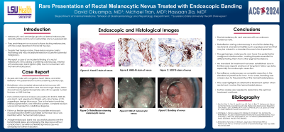
P0265 - Rare Presentation of Rectal Melanocytic Nevus Treated with Endoscopic Banding
Sunday, October 27, 2024
3:30 PM - 7:00 PM ET
Location: Exhibit Hall E

Has Audio

David Okuampa, MD
Louisiana State University Health
Shreveport, LA
Presenting Author(s)
David Okuampa, MD1, Michael Tran, MD1, Hassaan A. Zia, MD2
1Ochsner LSU Health, Shreveport, LA; 2LSU Health, Shreveport, LA
Introduction: Melanocytic nevi are benign growths of dermal melanocytes, typically solitary and found on extremities and sun-exposed skin. They are infrequent on mucosal surfaces lacking melanocytes, with few cases reported in the rectal mucosa. Despite their benign nature, these lesions require careful monitoring and may necessitate resection to prevent potential malignancy. Here, we report a case of an incidental finding of a rectal melanocytic nevus during a screening colonoscopy, treated with endoscopic band ligation (EBL) due to involvement with the hemorrhoidal plexus.
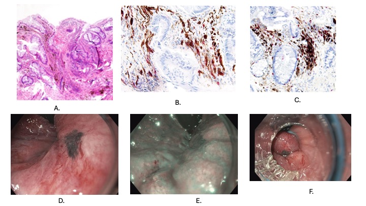
Case Description/Methods: 56-year-old male with congestive heart failure, atrial fibrillation who presented for routine screening colonoscopy. Retroflexion view revealed abnormal rectal mucosa with localized hyperpigmentation near the anal verge. Biopsy taken showed heavily pigmented spindle cells with low-grade nuclear features on H&E staining. Immunohistochemical analysis was positive for SOX10, HMB 45, and Mart1, and negative for PRAME, with a Ki-67 index of 1%, suggesting a benign blue nevus. Due to the lesion's small size, intense pigmentation, and infiltrative pattern, complete excision was recommended for a definitive diagnosis.
Follow-up flexible sigmoidoscopy for possible endoscopic mucosal resection (EMR) was halted as the blue nevus was identified within the hemorrhoidal plexus. Single endoscopic band was successfully placed over the hemorrhoidal plexus encompassing the blue nevus without complications. Surveillance flexible sigmoidoscopy was scheduled for further evaluation.
Discussion: Rectal melanocytic nevi are rare with an unknown incidence rate. Retroflexion during colonoscopy is crucial for detecting rectal and anal abnormalities such as polyps and nevi that may be missed in a standard forward-view inspection. Though benign, melanocytic nevi have the potential for malignant transformation, making biopsies essential for differentiating them from other pigmented lesions. No standard for treatment has been established due to limited case reports and lack of long-term follow-up data, especially for endoscopic resection. Surveillance colonoscopy or complete resection is the standard of practice for now. In our case banding was chosen due to proximity of the hemorrhoidal plexus. This case highlights an alternative treatment option when rectal nevi involve the hemorrhoidal plexus. Further studies are needed to determine the optimal treatment methods.

Disclosures:
David Okuampa, MD1, Michael Tran, MD1, Hassaan A. Zia, MD2. P0265 - Rare Presentation of Rectal Melanocytic Nevus Treated with Endoscopic Banding, ACG 2024 Annual Scientific Meeting Abstracts. Philadelphia, PA: American College of Gastroenterology.
1Ochsner LSU Health, Shreveport, LA; 2LSU Health, Shreveport, LA
Introduction: Melanocytic nevi are benign growths of dermal melanocytes, typically solitary and found on extremities and sun-exposed skin. They are infrequent on mucosal surfaces lacking melanocytes, with few cases reported in the rectal mucosa. Despite their benign nature, these lesions require careful monitoring and may necessitate resection to prevent potential malignancy. Here, we report a case of an incidental finding of a rectal melanocytic nevus during a screening colonoscopy, treated with endoscopic band ligation (EBL) due to involvement with the hemorrhoidal plexus.
Case Description/Methods: 56-year-old male with congestive heart failure, atrial fibrillation who presented for routine screening colonoscopy. Retroflexion view revealed abnormal rectal mucosa with localized hyperpigmentation near the anal verge. Biopsy taken showed heavily pigmented spindle cells with low-grade nuclear features on H&E staining. Immunohistochemical analysis was positive for SOX10, HMB 45, and Mart1, and negative for PRAME, with a Ki-67 index of 1%, suggesting a benign blue nevus. Due to the lesion's small size, intense pigmentation, and infiltrative pattern, complete excision was recommended for a definitive diagnosis.
Follow-up flexible sigmoidoscopy for possible endoscopic mucosal resection (EMR) was halted as the blue nevus was identified within the hemorrhoidal plexus. Single endoscopic band was successfully placed over the hemorrhoidal plexus encompassing the blue nevus without complications. Surveillance flexible sigmoidoscopy was scheduled for further evaluation.
Discussion: Rectal melanocytic nevi are rare with an unknown incidence rate. Retroflexion during colonoscopy is crucial for detecting rectal and anal abnormalities such as polyps and nevi that may be missed in a standard forward-view inspection. Though benign, melanocytic nevi have the potential for malignant transformation, making biopsies essential for differentiating them from other pigmented lesions. No standard for treatment has been established due to limited case reports and lack of long-term follow-up data, especially for endoscopic resection. Surveillance colonoscopy or complete resection is the standard of practice for now. In our case banding was chosen due to proximity of the hemorrhoidal plexus. This case highlights an alternative treatment option when rectal nevi involve the hemorrhoidal plexus. Further studies are needed to determine the optimal treatment methods.

Figure: A. H and E stain of nevus
B. HMB 45 stain of nevus
C. SOX10 stain of nevus
D. Retroflexion showing melanocytic nevus
E. NBI of melanocytic nevus
F. Banding of nevus
B. HMB 45 stain of nevus
C. SOX10 stain of nevus
D. Retroflexion showing melanocytic nevus
E. NBI of melanocytic nevus
F. Banding of nevus
Disclosures:
David Okuampa indicated no relevant financial relationships.
Michael Tran indicated no relevant financial relationships.
Hassaan A. Zia indicated no relevant financial relationships.
David Okuampa, MD1, Michael Tran, MD1, Hassaan A. Zia, MD2. P0265 - Rare Presentation of Rectal Melanocytic Nevus Treated with Endoscopic Banding, ACG 2024 Annual Scientific Meeting Abstracts. Philadelphia, PA: American College of Gastroenterology.
