Sunday Poster Session
Category: Colon
P0274 - An Unusual Case of Adult Intussuception Masquerading as an Ulcerated Colonic Mass
Sunday, October 27, 2024
3:30 PM - 7:00 PM ET
Location: Exhibit Hall E

Has Audio

Idowu Ajose, MD
University of Minnesota
Minneapolis, MN
Presenting Author(s)
Idowu Ajose, MD1, Taiwo Ajose, MD2, Bryant Megna, MD3, Justin Howard, MD2
1University of Minnesota, Minneapolis, MN; 2The University of Minnesota Medical School, Minneapolis, MN; 3University of Minnesota Medical Center, Minneapolis, MN
Introduction: Intussusception occurs rarely in adults. The presentation can involve a wide range of acute, intermittent,
and chronic symptoms, consequently making preoperative diagnosis challenging.
In contrast to pediatric intussusception, which is typically idiopathic, adult
intussusception (AI) is generally associated with an underlying cause, often some nature of mass. We describe an unusual case of an AI masquerading as a colonic mass.
Case Description/Methods: A 57-year-old male with a past medical history of major depressive disorder, constipation, internal hemorrhoids, and lipomas presented with intermittent loose stools and
abdominal cramping for two months. Further evaluation with a CT scan revealed findings suggestive of
colitis and a large intraluminal lipoma in the ascending colon.
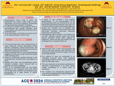
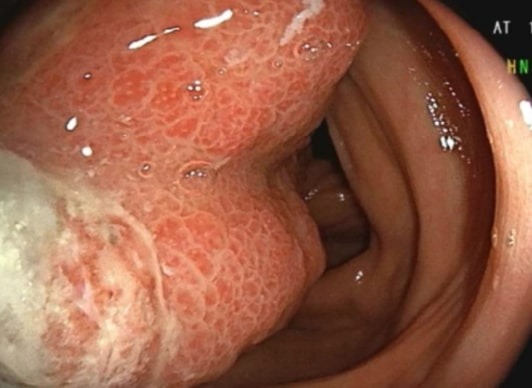
Follow up colonoscopy revealed ulceration in the ascending colon, associated with a mass [figures 1a &
1b]. Biopsies were taken, and histopathology reports indicated ulcerated colonic mucosa with no
dysplasia or invasive malignancy.
Prior to his scheduled follow up, the patient presented to the emergency room with severe (10/10) right
lower quadrant and suprapubic pain, accompanied by a large volume of painful hematochezia. A repeat
CT scan revealed a long segment (approximately 16 cm) intussusception of the cecum/ascending colon
and terminal ileum into the hepatic flexure/proximal transverse colon, with a 3.5 cm colonic lipoma
serving as the lead point (figure 2).
Surgery was consulted, and an emergent right hemicolectomy, resection of the terminal ileum with side-
to-side functional end-to-end ileocolonic anastomosis was performed. Histopathology report revealed a
cecal lipoma (5.8 cm) with overlying mucosal erosion and twelve reactive mesenteric lymph nodes,
negative for malignancy. Patient did well post-surgery and was discharged with an outpatient follow up.
Discussion: Approximately 52% of adult intussusceptions (AI) occur in the small intestine, while 38% (with 17%
specifically involving the ileocecal region) are found in the large intestine. The majority of cases are
attributable to an underlying structural lesion, with neoplasms accounting for 66% of colonic
intussusceptions and 30% of small bowel cases.
This case represents an uncommon presentation of ileocolonic intussusception in an adult patient,
initially manifesting as an ulcerated colonic mass on endoscopy. The condition was subsequently
diagnosed with cross sectional imaging and promptly managed with timely surgical intervention.

Disclosures:
Idowu Ajose, MD1, Taiwo Ajose, MD2, Bryant Megna, MD3, Justin Howard, MD2. P0274 - An Unusual Case of Adult Intussuception Masquerading as an Ulcerated Colonic Mass, ACG 2024 Annual Scientific Meeting Abstracts. Philadelphia, PA: American College of Gastroenterology.
1University of Minnesota, Minneapolis, MN; 2The University of Minnesota Medical School, Minneapolis, MN; 3University of Minnesota Medical Center, Minneapolis, MN
Introduction: Intussusception occurs rarely in adults. The presentation can involve a wide range of acute, intermittent,
and chronic symptoms, consequently making preoperative diagnosis challenging.
In contrast to pediatric intussusception, which is typically idiopathic, adult
intussusception (AI) is generally associated with an underlying cause, often some nature of mass. We describe an unusual case of an AI masquerading as a colonic mass.
Case Description/Methods: A 57-year-old male with a past medical history of major depressive disorder, constipation, internal hemorrhoids, and lipomas presented with intermittent loose stools and
abdominal cramping for two months. Further evaluation with a CT scan revealed findings suggestive of
colitis and a large intraluminal lipoma in the ascending colon.
Follow up colonoscopy revealed ulceration in the ascending colon, associated with a mass [figures 1a &
1b]. Biopsies were taken, and histopathology reports indicated ulcerated colonic mucosa with no
dysplasia or invasive malignancy.
Prior to his scheduled follow up, the patient presented to the emergency room with severe (10/10) right
lower quadrant and suprapubic pain, accompanied by a large volume of painful hematochezia. A repeat
CT scan revealed a long segment (approximately 16 cm) intussusception of the cecum/ascending colon
and terminal ileum into the hepatic flexure/proximal transverse colon, with a 3.5 cm colonic lipoma
serving as the lead point (figure 2).
Surgery was consulted, and an emergent right hemicolectomy, resection of the terminal ileum with side-
to-side functional end-to-end ileocolonic anastomosis was performed. Histopathology report revealed a
cecal lipoma (5.8 cm) with overlying mucosal erosion and twelve reactive mesenteric lymph nodes,
negative for malignancy. Patient did well post-surgery and was discharged with an outpatient follow up.
Discussion: Approximately 52% of adult intussusceptions (AI) occur in the small intestine, while 38% (with 17%
specifically involving the ileocecal region) are found in the large intestine. The majority of cases are
attributable to an underlying structural lesion, with neoplasms accounting for 66% of colonic
intussusceptions and 30% of small bowel cases.
This case represents an uncommon presentation of ileocolonic intussusception in an adult patient,
initially manifesting as an ulcerated colonic mass on endoscopy. The condition was subsequently
diagnosed with cross sectional imaging and promptly managed with timely surgical intervention.

Figure: Figure 1A
Disclosures:
Idowu Ajose indicated no relevant financial relationships.
Taiwo Ajose indicated no relevant financial relationships.
Bryant Megna indicated no relevant financial relationships.
Justin Howard indicated no relevant financial relationships.
Idowu Ajose, MD1, Taiwo Ajose, MD2, Bryant Megna, MD3, Justin Howard, MD2. P0274 - An Unusual Case of Adult Intussuception Masquerading as an Ulcerated Colonic Mass, ACG 2024 Annual Scientific Meeting Abstracts. Philadelphia, PA: American College of Gastroenterology.
