Sunday Poster Session
Category: Biliary/Pancreas
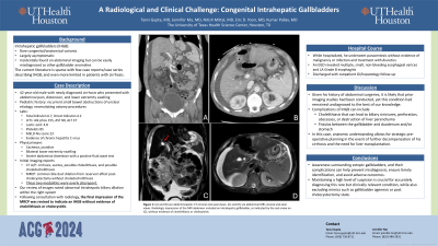
P0196 - A Radiological and Clinical Challenge: Congenital Intrahepatic Gallbladders
Sunday, October 27, 2024
3:30 PM - 7:00 PM ET
Location: Exhibit Hall E

Has Audio

Tanvi Gupta, MD
University of Texas Health, McGovern Medical School
Houston, TX
Presenting Author(s)
Tanvi Gupta, MD1, Jennifer Ma, MD1, Nitish Mittal, MD1, Eric D. Yoon, MD2, Kumar Pallav, MD1
1University of Texas Health, McGovern Medical School, Houston, TX; 2University of Texas at Houston, Houston, TX
Introduction: Intrahepatic gallbladders (IHGB) are rare congenital variants, largely asymptomatic, and incidentally found on abdominal imaging but can be easily misdiagnosed as other gallbladder anomalies. The current literature is sparse with few case reports describing IHGB, and even more limited in patients with cirrhosis.
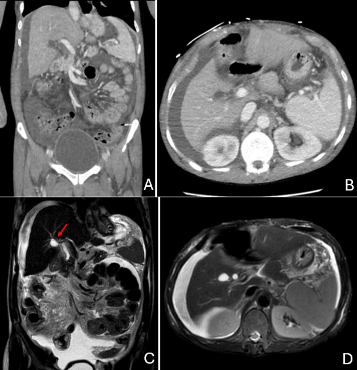
Case Description/Methods: We report a case of a 42-year-old male with newly diagnosed cirrhosis, who presented with abdominal pain, distension, and lower extremity swelling. His pediatric history included recurrent small bowel obstructions of unclear etiology, necessitating ostomy procedures. Laboratory abnormalities included elevated transaminases, hyperbilirubinemia, thrombocytopenia, lactic acidosis, and evidence of chronic hepatitis C virus. Pertinent physical exam findings included cachexia, jaundice, bilateral lower extremity swelling, and severe abdominal distention with a positive fluid wave test. Abdominopelvic CT scan showed cirrhosis, ascites, possible cholelithiasis, and possible choledocholithiasis. A subsequent MRCP reported common bile duct dilation from reservoir effect post-cholecystectomy without choledocholithiasis. The two modalities were overly discrepant. Our review of images noted abnormal intrahepatic biliary dilation within the right system. Following consultation with radiology, the final impression from MRCP was revised to indicate an IHGB without evidence of cholelithiasis or cholecystitis. Given his history of abdominal surgeries, it is likely that prior imaging studies had been conducted, yet this condition had remained undiagnosed to the best of our knowledge. While hospitalized, he underwent paracentesis without evidence of malignancy or infection and treatment with diuretics. An EGD revealed non-bleeding esophageal varices and LA Grade B esophagitis. He was discharged with outpatient follow-up.
Discussion: Maintaining a high level of suspicion is crucial for accurately diagnosing this rare but clinically relevant condition, while also ruling out mimics such as gallbladder agenesis or post cholecystectomy state. Complications of IHGB can include cholelithiasis or fistulas between the gallbladder and duodenum and/or stomach. In this case, anatomic understanding allows for strategic pre-operative planning in the event of further decompensation of his cirrhosis and the need for liver transplantation. Awareness surrounding ectopic gallbladders and their complications can help prevent misdiagnosis, ensure timely identification, and avoid adverse outcomes.

Disclosures:
Tanvi Gupta, MD1, Jennifer Ma, MD1, Nitish Mittal, MD1, Eric D. Yoon, MD2, Kumar Pallav, MD1. P0196 - A Radiological and Clinical Challenge: Congenital Intrahepatic Gallbladders, ACG 2024 Annual Scientific Meeting Abstracts. Philadelphia, PA: American College of Gastroenterology.
1University of Texas Health, McGovern Medical School, Houston, TX; 2University of Texas at Houston, Houston, TX
Introduction: Intrahepatic gallbladders (IHGB) are rare congenital variants, largely asymptomatic, and incidentally found on abdominal imaging but can be easily misdiagnosed as other gallbladder anomalies. The current literature is sparse with few case reports describing IHGB, and even more limited in patients with cirrhosis.
Case Description/Methods: We report a case of a 42-year-old male with newly diagnosed cirrhosis, who presented with abdominal pain, distension, and lower extremity swelling. His pediatric history included recurrent small bowel obstructions of unclear etiology, necessitating ostomy procedures. Laboratory abnormalities included elevated transaminases, hyperbilirubinemia, thrombocytopenia, lactic acidosis, and evidence of chronic hepatitis C virus. Pertinent physical exam findings included cachexia, jaundice, bilateral lower extremity swelling, and severe abdominal distention with a positive fluid wave test. Abdominopelvic CT scan showed cirrhosis, ascites, possible cholelithiasis, and possible choledocholithiasis. A subsequent MRCP reported common bile duct dilation from reservoir effect post-cholecystectomy without choledocholithiasis. The two modalities were overly discrepant. Our review of images noted abnormal intrahepatic biliary dilation within the right system. Following consultation with radiology, the final impression from MRCP was revised to indicate an IHGB without evidence of cholelithiasis or cholecystitis. Given his history of abdominal surgeries, it is likely that prior imaging studies had been conducted, yet this condition had remained undiagnosed to the best of our knowledge. While hospitalized, he underwent paracentesis without evidence of malignancy or infection and treatment with diuretics. An EGD revealed non-bleeding esophageal varices and LA Grade B esophagitis. He was discharged with outpatient follow-up.
Discussion: Maintaining a high level of suspicion is crucial for accurately diagnosing this rare but clinically relevant condition, while also ruling out mimics such as gallbladder agenesis or post cholecystectomy state. Complications of IHGB can include cholelithiasis or fistulas between the gallbladder and duodenum and/or stomach. In this case, anatomic understanding allows for strategic pre-operative planning in the event of further decompensation of his cirrhosis and the need for liver transplantation. Awareness surrounding ectopic gallbladders and their complications can help prevent misdiagnosis, ensure timely identification, and avoid adverse outcomes.

Figure: Figure 1: (A) Abdominopelvic CT coronal view, (B) Abdominopelvic CT axial view, (C) Abdominal MRI coronal view, (D) Abdominal MRI axial view. Radiologic impression of the MRI abdomen included an intrahepatic gallbladder, as indicated by the red arrow on (C), without evidence of cholelithiasis or cholecystitis.
Disclosures:
Tanvi Gupta indicated no relevant financial relationships.
Jennifer Ma indicated no relevant financial relationships.
Nitish Mittal indicated no relevant financial relationships.
Eric Yoon indicated no relevant financial relationships.
Kumar Pallav indicated no relevant financial relationships.
Tanvi Gupta, MD1, Jennifer Ma, MD1, Nitish Mittal, MD1, Eric D. Yoon, MD2, Kumar Pallav, MD1. P0196 - A Radiological and Clinical Challenge: Congenital Intrahepatic Gallbladders, ACG 2024 Annual Scientific Meeting Abstracts. Philadelphia, PA: American College of Gastroenterology.
