Sunday Poster Session
Category: Biliary/Pancreas
P0105 - Rare Presentation of Eosinophilic Cholangitis in a 32-Year-Old Male: A Case Report
Sunday, October 27, 2024
3:30 PM - 7:00 PM ET
Location: Exhibit Hall E

Has Audio

Himsikhar Khataniar, MD
Allegheny General Hospital
Pittsburgh, PA
Presenting Author(s)
Award: Presidential Poster Award
Himsikhar Khataniar, MD1, Molly Ruiz, BS, MS2, Ashni Dharia, MD1, Sheena Mago, DO3, Muhammad Ali Butt, MD1, Abhijit Kulkarni, MD4
1Allegheny General Hospital, Pittsburgh, PA; 2Drexel University College of Medicine, Pittsburgh, PA; 3Allegheny Health Network, Pittsburgh, PA; 4Allegheny Center for Digestive Health, Pittsburgh, PA
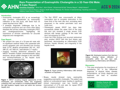
Introduction: Eosinophilic cholangitis (EC) is an exceedingly rare condition characterized by eosinophilic infiltration of the bile ducts, with fewer than 30 cases reported since 1980. It presents diagnostic challenges due to its nonspecific symptoms and can mimic other biliary diseases like primary sclerosing cholangitis (PSC) and cholangiocarcinoma, highlighting the importance of clinician awareness for accurate diagnosis and management.
Case Description/Methods: We present the case of a 32-year-old male with no significant medical history who presented with severe epigastric pain and elevated liver function tests (LFTs: alkaline phosphatase 263 U/L, AST 286 U/L, ALT 659 U/L, total bilirubin 2.7 mg/dL). Imaging, including abdominal ultrasound and MRCP, revealed multiple intra/extrahepatic biliary strictures, abrupt caliber changes in the mid-CBD, and choledocholithiasis in the hepatic ducts suggestive of underlying PSC.
The first ERCP was unsuccessful at biliary cannulation due to complete obstruction in the lower third of the main bile duct, prompting stent placement in the ventral pancreatic duct and cystic duct. Subsequent repeat ERCP two days later successfully dilated the upper third of the main bile duct and revealed a single severe CHD stricture with benign cytology of the main bile duct, but polysomy on FISH. Given the FISH findings and recurrent abdominal pain, another ERCP with bile duct biopsy was performed, demonstrating an inflammatory CHD stricture, hepatic lithiasis, and irregularity in the hepatic ducts. Biopsy results showed many eosinophils, suggesting eosinophilic cholangitis. The patient was started on corticosteroid taper, leading to significant clinical improvement over two months and improvement of LFTs and plan for repeat ERCP after completion of steroids.
Discussion: This case emphasizes the importance of considering EC in the differential diagnosis of atypical biliary disease presentations, as timely diagnosis and treatment can lead to excellent outcomes.

Disclosures:
Himsikhar Khataniar, MD1, Molly Ruiz, BS, MS2, Ashni Dharia, MD1, Sheena Mago, DO3, Muhammad Ali Butt, MD1, Abhijit Kulkarni, MD4. P0105 - Rare Presentation of Eosinophilic Cholangitis in a 32-Year-Old Male: A Case Report, ACG 2024 Annual Scientific Meeting Abstracts. Philadelphia, PA: American College of Gastroenterology.
Himsikhar Khataniar, MD1, Molly Ruiz, BS, MS2, Ashni Dharia, MD1, Sheena Mago, DO3, Muhammad Ali Butt, MD1, Abhijit Kulkarni, MD4
1Allegheny General Hospital, Pittsburgh, PA; 2Drexel University College of Medicine, Pittsburgh, PA; 3Allegheny Health Network, Pittsburgh, PA; 4Allegheny Center for Digestive Health, Pittsburgh, PA
Introduction: Eosinophilic cholangitis (EC) is an exceedingly rare condition characterized by eosinophilic infiltration of the bile ducts, with fewer than 30 cases reported since 1980. It presents diagnostic challenges due to its nonspecific symptoms and can mimic other biliary diseases like primary sclerosing cholangitis (PSC) and cholangiocarcinoma, highlighting the importance of clinician awareness for accurate diagnosis and management.
Case Description/Methods: We present the case of a 32-year-old male with no significant medical history who presented with severe epigastric pain and elevated liver function tests (LFTs: alkaline phosphatase 263 U/L, AST 286 U/L, ALT 659 U/L, total bilirubin 2.7 mg/dL). Imaging, including abdominal ultrasound and MRCP, revealed multiple intra/extrahepatic biliary strictures, abrupt caliber changes in the mid-CBD, and choledocholithiasis in the hepatic ducts suggestive of underlying PSC.
The first ERCP was unsuccessful at biliary cannulation due to complete obstruction in the lower third of the main bile duct, prompting stent placement in the ventral pancreatic duct and cystic duct. Subsequent repeat ERCP two days later successfully dilated the upper third of the main bile duct and revealed a single severe CHD stricture with benign cytology of the main bile duct, but polysomy on FISH. Given the FISH findings and recurrent abdominal pain, another ERCP with bile duct biopsy was performed, demonstrating an inflammatory CHD stricture, hepatic lithiasis, and irregularity in the hepatic ducts. Biopsy results showed many eosinophils, suggesting eosinophilic cholangitis. The patient was started on corticosteroid taper, leading to significant clinical improvement over two months and improvement of LFTs and plan for repeat ERCP after completion of steroids.
Discussion: This case emphasizes the importance of considering EC in the differential diagnosis of atypical biliary disease presentations, as timely diagnosis and treatment can lead to excellent outcomes.

Figure: Figure 1: MRCP revealing irregular appearance of the intrahepatic and extrahepatic biliary tree suggesting underlying PSC and evidence of choledocholithiasis within the right posterior hepatic ducts and within the common hepatic duct.
Figure 2: Inflammatory CBD stricture visualized via SpyGlass
Figure 2: Inflammatory CBD stricture visualized via SpyGlass
Disclosures:
Himsikhar Khataniar indicated no relevant financial relationships.
Molly Ruiz indicated no relevant financial relationships.
Ashni Dharia indicated no relevant financial relationships.
Sheena Mago indicated no relevant financial relationships.
Muhammad Ali Butt indicated no relevant financial relationships.
Abhijit Kulkarni indicated no relevant financial relationships.
Himsikhar Khataniar, MD1, Molly Ruiz, BS, MS2, Ashni Dharia, MD1, Sheena Mago, DO3, Muhammad Ali Butt, MD1, Abhijit Kulkarni, MD4. P0105 - Rare Presentation of Eosinophilic Cholangitis in a 32-Year-Old Male: A Case Report, ACG 2024 Annual Scientific Meeting Abstracts. Philadelphia, PA: American College of Gastroenterology.

