Sunday Poster Session
Category: Biliary/Pancreas
P0107 - Primary Pancreatic Follicular Lymphoma: Insights from a Rare Case
Sunday, October 27, 2024
3:30 PM - 7:00 PM ET
Location: Exhibit Hall E

Has Audio

Jose I. De Arrigunaga, DO
University of Texas Health San Antonio
San Antonio, TX
Presenting Author(s)
Jose I.. De Arrigunaga, DO1, Umar Siddiqui, DO1, Apurva Bonde, MD1, Hari Sayana, MBBS, MD2
1University of Texas Health San Antonio, San Antonio, TX; 2University Hospital, San Antonio, TX
Introduction: Primary pancreatic lymphomas are extremely unusual; they account for only 0.1-0.5% of malignant lymphomas and 0.2% of primary pancreatic tumors. The two most common types include diffuse large B-cell lymphoma and follicular lymphoma (FL). In the past 30 years, there have only been 31 cases of primary pancreatic follicular lymphoma (PPFL) reported in the literature. We present a case of a middle-aged male with back pain and weight loss who was found to have PPFL.
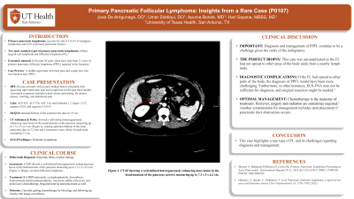
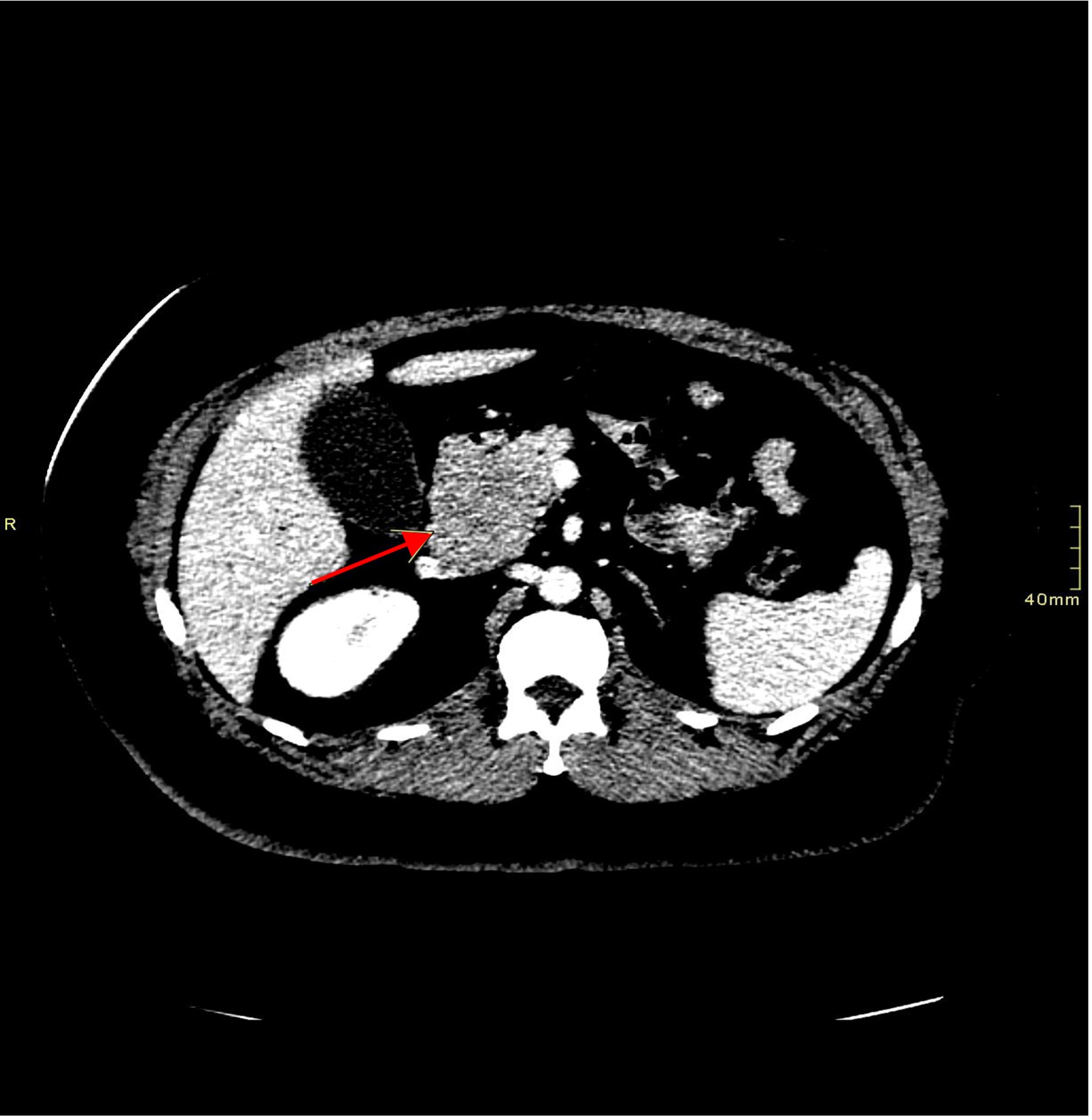
Case Description/Methods: A 48-year-old male with no past medical history presented with squeezing right-sided back pain and weight loss for the past three months. Associated symptoms included scleral icterus and itching. He denied nausea, vomiting, and abdominal pain. Labs were significant for alkaline phosphatase 455, alanine aminotransferase 276, aspartate aminotransferase 116, total bilirubin 1.2, lipase 1,337, negative carcinoembryonic antigen, and negative carbohydrate antigen 19-9. Right upper quadrant ultrasound showed dilation of the common bile duct at 1.9 cm. Computed tomography of the abdomen and pelvis (CTAP) showed a well-defined heterogeneously enhancing mass lesion in the head/uncinate of the pancreas measuring up to 7.3 x 5 x 6.2 cm (Figure 1), causing upstream dilation of the main pancreatic duct at 3.2 mm and a mesenteric mass, likely a lymph node, measuring 2.3 cm. Endoscopic ultrasound-guided fine-needle aspiration (EUS-FNA) was performed, and a biopsy was obtained. Biopsy of the mesenteric lymph node was also obtained. Endoscopic retrograde cholangiopancreatography was performed, and biliary sphincterotomy was done along with stent placement. The pathology report showed FL on both biopsies. The patient was discharged with follow-up appointments for medical oncology, surgical oncology, and advanced endoscopy.
Discussion: Diagnosis and management of PPFL continue to be a challenge given the rarity of the malignancy. Diagnostically, this case was uncomplicated as the FL had not spread to other areas of the body aside from a nearby lymph node. If the FL had spread to other parts of the body, the diagnosis of PPFL would have been more challenging. Furthermore, in other instances, EUS-FNA may not be sufficient for diagnosis, and surgical resection might be needed. Treatment for PPFL includes chemotherapy, surgery, and radiation. Another consideration for management includes stent placement if pancreatic duct obstruction occurs. This case highlights a rare type of FL and its challenges regarding diagnosis and management.

Disclosures:
Jose I.. De Arrigunaga, DO1, Umar Siddiqui, DO1, Apurva Bonde, MD1, Hari Sayana, MBBS, MD2. P0107 - Primary Pancreatic Follicular Lymphoma: Insights from a Rare Case, ACG 2024 Annual Scientific Meeting Abstracts. Philadelphia, PA: American College of Gastroenterology.
1University of Texas Health San Antonio, San Antonio, TX; 2University Hospital, San Antonio, TX
Introduction: Primary pancreatic lymphomas are extremely unusual; they account for only 0.1-0.5% of malignant lymphomas and 0.2% of primary pancreatic tumors. The two most common types include diffuse large B-cell lymphoma and follicular lymphoma (FL). In the past 30 years, there have only been 31 cases of primary pancreatic follicular lymphoma (PPFL) reported in the literature. We present a case of a middle-aged male with back pain and weight loss who was found to have PPFL.
Case Description/Methods: A 48-year-old male with no past medical history presented with squeezing right-sided back pain and weight loss for the past three months. Associated symptoms included scleral icterus and itching. He denied nausea, vomiting, and abdominal pain. Labs were significant for alkaline phosphatase 455, alanine aminotransferase 276, aspartate aminotransferase 116, total bilirubin 1.2, lipase 1,337, negative carcinoembryonic antigen, and negative carbohydrate antigen 19-9. Right upper quadrant ultrasound showed dilation of the common bile duct at 1.9 cm. Computed tomography of the abdomen and pelvis (CTAP) showed a well-defined heterogeneously enhancing mass lesion in the head/uncinate of the pancreas measuring up to 7.3 x 5 x 6.2 cm (Figure 1), causing upstream dilation of the main pancreatic duct at 3.2 mm and a mesenteric mass, likely a lymph node, measuring 2.3 cm. Endoscopic ultrasound-guided fine-needle aspiration (EUS-FNA) was performed, and a biopsy was obtained. Biopsy of the mesenteric lymph node was also obtained. Endoscopic retrograde cholangiopancreatography was performed, and biliary sphincterotomy was done along with stent placement. The pathology report showed FL on both biopsies. The patient was discharged with follow-up appointments for medical oncology, surgical oncology, and advanced endoscopy.
Discussion: Diagnosis and management of PPFL continue to be a challenge given the rarity of the malignancy. Diagnostically, this case was uncomplicated as the FL had not spread to other areas of the body aside from a nearby lymph node. If the FL had spread to other parts of the body, the diagnosis of PPFL would have been more challenging. Furthermore, in other instances, EUS-FNA may not be sufficient for diagnosis, and surgical resection might be needed. Treatment for PPFL includes chemotherapy, surgery, and radiation. Another consideration for management includes stent placement if pancreatic duct obstruction occurs. This case highlights a rare type of FL and its challenges regarding diagnosis and management.

Figure: Figure 1. CTAP showing a well-defined heterogeneously enhancing mass lesion in the head/uncinate of the pancreas (arrow) measuring up to 7.3 x 5 x 6.2 cm.
Disclosures:
Jose De Arrigunaga indicated no relevant financial relationships.
Umar Siddiqui indicated no relevant financial relationships.
Apurva Bonde indicated no relevant financial relationships.
Hari Sayana indicated no relevant financial relationships.
Jose I.. De Arrigunaga, DO1, Umar Siddiqui, DO1, Apurva Bonde, MD1, Hari Sayana, MBBS, MD2. P0107 - Primary Pancreatic Follicular Lymphoma: Insights from a Rare Case, ACG 2024 Annual Scientific Meeting Abstracts. Philadelphia, PA: American College of Gastroenterology.
