Sunday Poster Session
Category: Biliary/Pancreas
P0111 - Obstructive Enigma: A Clinical Insight Into the Bouveret Syndrome
Sunday, October 27, 2024
3:30 PM - 7:00 PM ET
Location: Exhibit Hall E

Has Audio

Pujitha Vallivedu Chennakesavulu, MD, MBBS
Quinnipiac University Frank H. Netter MD School of Medicine / St. Vincent's Medical Center
Bridgeport, CT
Presenting Author(s)
Pujitha Vallivedu Chennakesavulu, MD, MBBS, Sushrut Ingawale, MD, MBBS, Prabin Sharma, MD
Quinnipiac University Frank H. Netter MD School of Medicine / St. Vincent's Medical Center, Bridgeport, CT
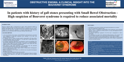
Introduction: Bouveret syndrome (BS) is characterized by gastric outlet obstruction (GOO) by gallstones passing through the biliary-enteric fistula. It accounts for 1-3 % of all patients with gallstone ileus. Our patient had BS with choledocho-duodenal fistula, managed by the consecutive procedure of emergent laparoscopy with obstructed gallstone retrieval followed by ERCP with stone extraction from the fistula.
Case Description/Methods: A 76-year-old female with a history of atrial fibrillation and hypertension, presented with complaints of right-sided abdominal pain radiating to the back, and nausea/vomiting for 3 days. CT abdomen with IV contrast revealed extensive pneumobilia with large caliber choledochoduodenal fistula and impacted 3.3 cm gall stone in distal small bowel consistent with GOO characterizing BS. An emergent exploratory laparotomy was performed to retrieve the gallstone. She had persistent abdominal pain post-procedure with concerns of choledocholithiasis related to fistula, MRCP was done revealing a gallstone in the common bile duct (CBD) at the level of a choledocho-duodenal fistula. EUS showed a large stone measuring 2 cm protruding from the presumed choledochoduodenal fistula proximal to the inflamed ampulla. This was retrieved using a balloon-tipped catheter. Intraprocedural cholangiogram showed a CBD dilation of 10 mm. Biliary cannulation via native papilla was deferred since the patient had adequate drainage via the fistula. The patient improved symptomatically post-procedure. A repeat abdominal CT scan 1 month later showed a persistent fistula, but further procedures were deferred as the patient was asymptomatic and had multiple comorbidities.
Discussion: Cholelithiasis is prevalent worldwide and atypical presentations are common. One rare complication is gallstone ileus. Rigler’s triad is a constellation of pneumobilia, bowel obstruction, and an aberrant gallstone, which is suggestive of BS in 40-50 % of cases and diagnosed with CT abdomen with contrast. An emergent laparoscopic approach is the first step in management to relieve obstruction. Few published case reports have performed cholecystectomy, stone retrieval, and closure of fistula within the same surgical approach but had higher morbidity. The procedure should be tailored individually; thus, a high suspicion of BS is needed in patients presenting with small bowel obstruction with a history of gallstones to decrease associated mortality.

Disclosures:
Pujitha Vallivedu Chennakesavulu, MD, MBBS, Sushrut Ingawale, MD, MBBS, Prabin Sharma, MD. P0111 - Obstructive Enigma: A Clinical Insight Into the Bouveret Syndrome, ACG 2024 Annual Scientific Meeting Abstracts. Philadelphia, PA: American College of Gastroenterology.
Quinnipiac University Frank H. Netter MD School of Medicine / St. Vincent's Medical Center, Bridgeport, CT
Introduction: Bouveret syndrome (BS) is characterized by gastric outlet obstruction (GOO) by gallstones passing through the biliary-enteric fistula. It accounts for 1-3 % of all patients with gallstone ileus. Our patient had BS with choledocho-duodenal fistula, managed by the consecutive procedure of emergent laparoscopy with obstructed gallstone retrieval followed by ERCP with stone extraction from the fistula.
Case Description/Methods: A 76-year-old female with a history of atrial fibrillation and hypertension, presented with complaints of right-sided abdominal pain radiating to the back, and nausea/vomiting for 3 days. CT abdomen with IV contrast revealed extensive pneumobilia with large caliber choledochoduodenal fistula and impacted 3.3 cm gall stone in distal small bowel consistent with GOO characterizing BS. An emergent exploratory laparotomy was performed to retrieve the gallstone. She had persistent abdominal pain post-procedure with concerns of choledocholithiasis related to fistula, MRCP was done revealing a gallstone in the common bile duct (CBD) at the level of a choledocho-duodenal fistula. EUS showed a large stone measuring 2 cm protruding from the presumed choledochoduodenal fistula proximal to the inflamed ampulla. This was retrieved using a balloon-tipped catheter. Intraprocedural cholangiogram showed a CBD dilation of 10 mm. Biliary cannulation via native papilla was deferred since the patient had adequate drainage via the fistula. The patient improved symptomatically post-procedure. A repeat abdominal CT scan 1 month later showed a persistent fistula, but further procedures were deferred as the patient was asymptomatic and had multiple comorbidities.
Discussion: Cholelithiasis is prevalent worldwide and atypical presentations are common. One rare complication is gallstone ileus. Rigler’s triad is a constellation of pneumobilia, bowel obstruction, and an aberrant gallstone, which is suggestive of BS in 40-50 % of cases and diagnosed with CT abdomen with contrast. An emergent laparoscopic approach is the first step in management to relieve obstruction. Few published case reports have performed cholecystectomy, stone retrieval, and closure of fistula within the same surgical approach but had higher morbidity. The procedure should be tailored individually; thus, a high suspicion of BS is needed in patients presenting with small bowel obstruction with a history of gallstones to decrease associated mortality.

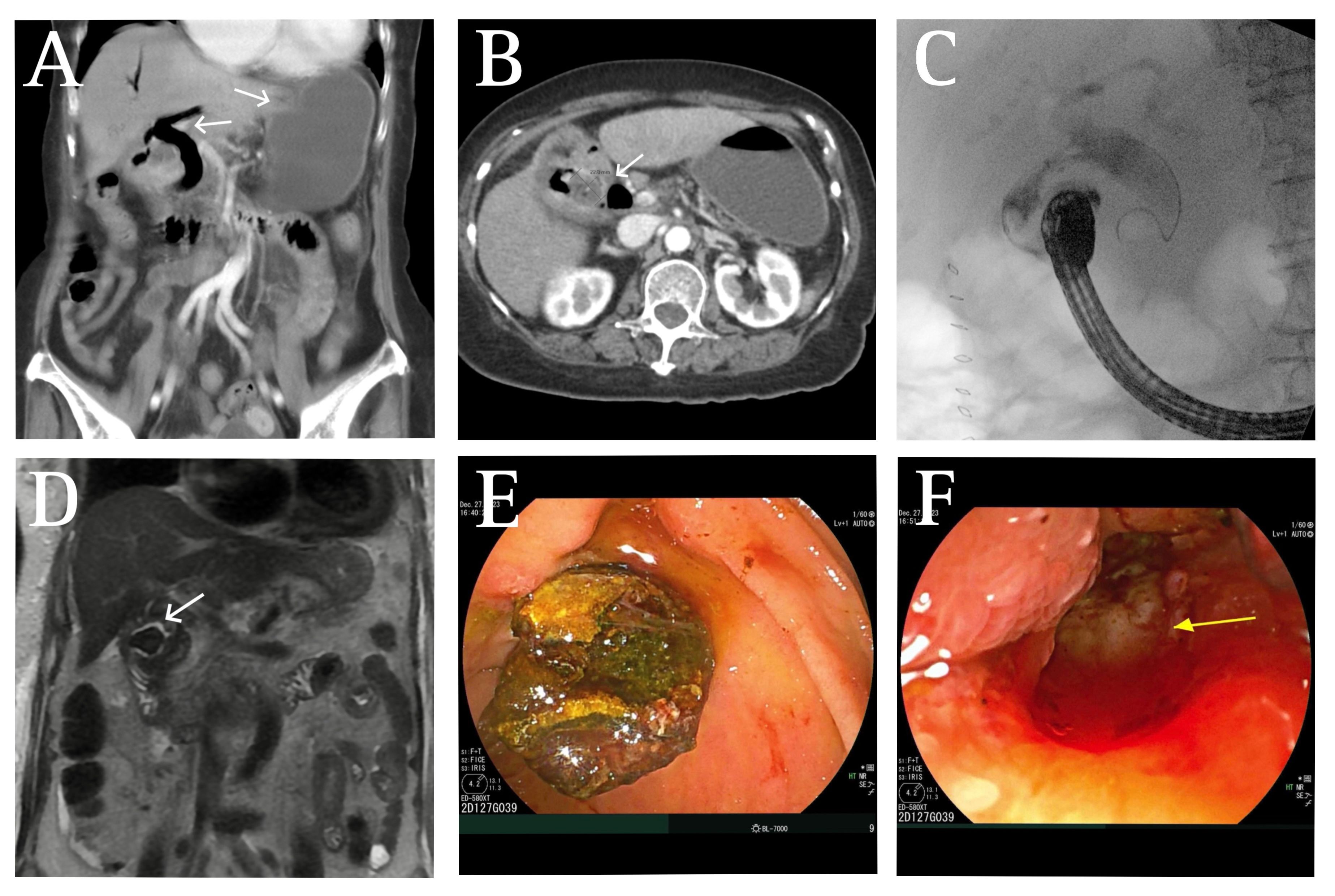
Figure: Image A: CT abdomen with IV contrast showing extensive pneumobilia and gastric outlet obstruction,
Image B: 2.3 cm rounded hypoattenuating structure representing a noncalcified gallstone near choledocho-duodenal fistula in CT abdomen with IV contrast,
Image C: ERCP - Fluoroscopy showing dilated common bile duct (CBD) with dye leaking into bowel loop indicating a fistula,
Image D: MRCP showing gall stone in the region of CBD/duodenum with improved pneumobilia and gaseous distention,
Image E: Duodenoscope showing gallstone extruding from choledocho-duodenal fistula proximal to ampulla,
Image F: Endoscopic view of choledochoduodenal fistula proximal to the ampulla after gallstone extraction
Disclosures:
Pujitha Vallivedu Chennakesavulu indicated no relevant financial relationships.
Sushrut Ingawale indicated no relevant financial relationships.
Prabin Sharma indicated no relevant financial relationships.
Pujitha Vallivedu Chennakesavulu, MD, MBBS, Sushrut Ingawale, MD, MBBS, Prabin Sharma, MD. P0111 - Obstructive Enigma: A Clinical Insight Into the Bouveret Syndrome, ACG 2024 Annual Scientific Meeting Abstracts. Philadelphia, PA: American College of Gastroenterology.
