Sunday Poster Session
Category: Biliary/Pancreas
P0118 - Bilothorax: A Rare Cause of Exudative Pleural Effusion in a Patient with Metastatic Colorectal Carcinoma
Sunday, October 27, 2024
3:30 PM - 7:00 PM ET
Location: Exhibit Hall E

Has Audio

Shan Xue, MD
Dartmouth Hitchcock Medical Center
Lebanon, NH
Presenting Author(s)
Shan Xue, MD, David Feller-Kopman, MD
Dartmouth Hitchcock Medical Center, Lebanon, NH
Introduction: Bilious pleural effusion, or bilothorax, is a rare cause of exudative effusion, characterized as bile in the pleural fluid. Here we describe a case of bilothorax in a patient with complicated biliary anatomy.
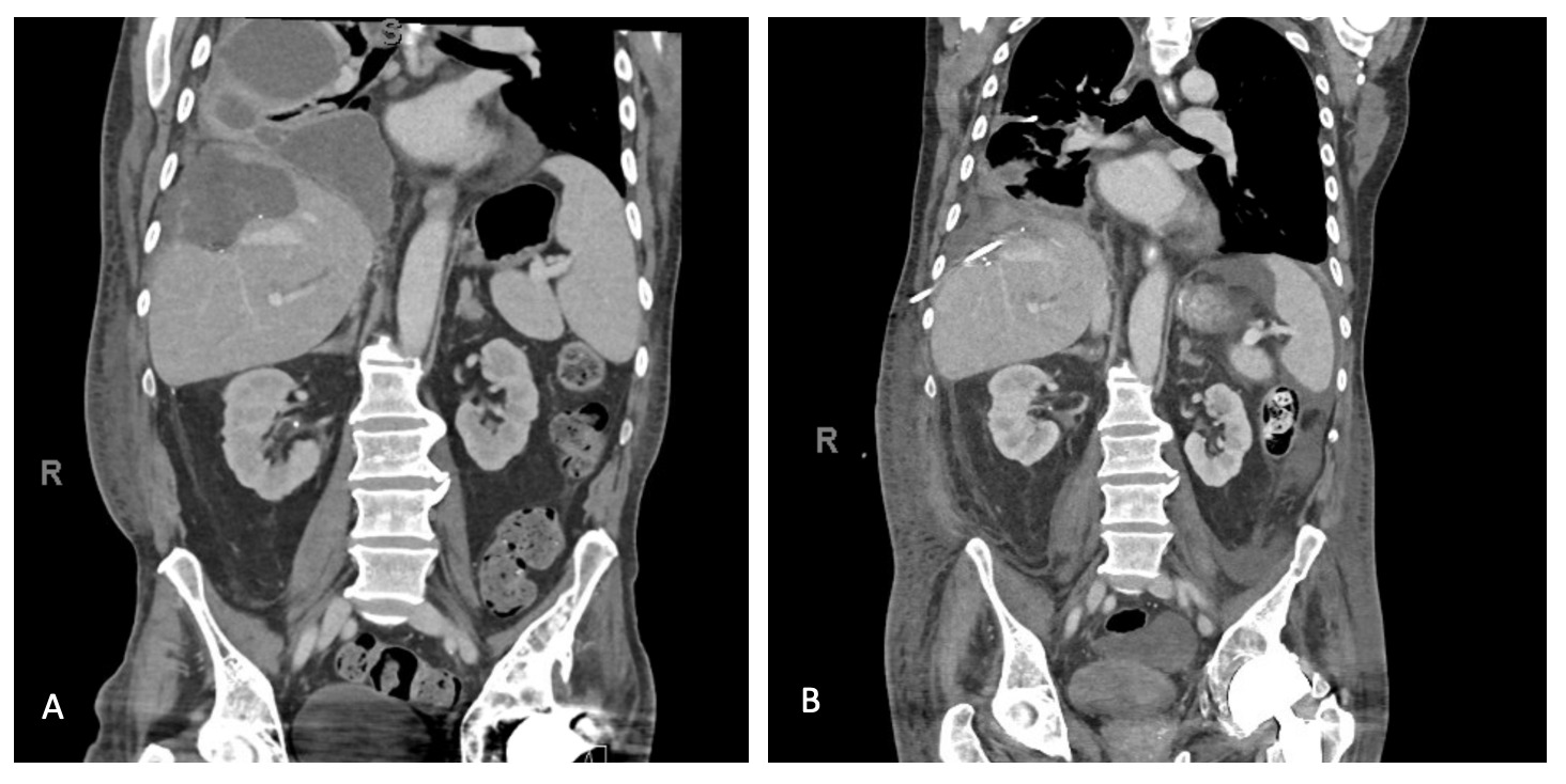
Case Description/Methods: A 62-year-old male patient presented to the hospital for evaluation of abdominal pain and shortness of breath. His medical history is notable for metastatic colorectal carcinoma to the liver status post right partial hepatectomy, complicated by malignant biliary obstruction requiring stent placement. He was also hospitalized 4 months prior for ascending cholangitis requiring biliary drain. Workup for his current presentation included a CT abdomen and pelvis which showed a large loculated right hydropneumothorax and a fluid collection at the partial R hepatectomy site abutting the undersurface of the R hemidiaphragm.
Pleural fluid studies were remarkable for WBC count 185,925 mcl, LDH >2,500 unit/L, proteins 2.5 g/dL, total bilirubin 5.6 mg/dL, and glucose < 2 mg/dL. His serum total bilirubin was 2.8 mg/dL, with a pleural bilirubin to serum bilirubin ratio of 2, suggestive of the diagnosis of bilothorax. He was started on broad-spectrum antibiotics for the concomitant empyema. Surgical teams were consulted for potential pleural/biliary communication but noted no role of surgical interventions.
Discussion: The most common etiologies of bilothorax include: biliary obstruction, diaphragmatic trauma or perforation, extension of hepatic infections or subphrenic abscesses, and iatrogenic complications of percutaneous procedures.1 The diagnosis of bilothorax requires a high index of suspicion, along with imaging demonstrating an effusion and compatible pleural fluid studies.2 Studies have reported that the most specific test for diagnosis is the ratio of pleural total bilirubin to serum total bilirubin of greater than 1, however, recent study by Saraya et al. have suggested that the conventional ratio is noted to be only 76.9% sensitive.1,3 While not required for diagnosis, demonstration of a biliary-pleural fistula on imaging would be useful for both evaluation and management.1
References:

Disclosures:
Shan Xue, MD, David Feller-Kopman, MD. P0118 - Bilothorax: A Rare Cause of Exudative Pleural Effusion in a Patient with Metastatic Colorectal Carcinoma, ACG 2024 Annual Scientific Meeting Abstracts. Philadelphia, PA: American College of Gastroenterology.
Dartmouth Hitchcock Medical Center, Lebanon, NH
Introduction: Bilious pleural effusion, or bilothorax, is a rare cause of exudative effusion, characterized as bile in the pleural fluid. Here we describe a case of bilothorax in a patient with complicated biliary anatomy.
Case Description/Methods: A 62-year-old male patient presented to the hospital for evaluation of abdominal pain and shortness of breath. His medical history is notable for metastatic colorectal carcinoma to the liver status post right partial hepatectomy, complicated by malignant biliary obstruction requiring stent placement. He was also hospitalized 4 months prior for ascending cholangitis requiring biliary drain. Workup for his current presentation included a CT abdomen and pelvis which showed a large loculated right hydropneumothorax and a fluid collection at the partial R hepatectomy site abutting the undersurface of the R hemidiaphragm.
Pleural fluid studies were remarkable for WBC count 185,925 mcl, LDH >2,500 unit/L, proteins 2.5 g/dL, total bilirubin 5.6 mg/dL, and glucose < 2 mg/dL. His serum total bilirubin was 2.8 mg/dL, with a pleural bilirubin to serum bilirubin ratio of 2, suggestive of the diagnosis of bilothorax. He was started on broad-spectrum antibiotics for the concomitant empyema. Surgical teams were consulted for potential pleural/biliary communication but noted no role of surgical interventions.
Discussion: The most common etiologies of bilothorax include: biliary obstruction, diaphragmatic trauma or perforation, extension of hepatic infections or subphrenic abscesses, and iatrogenic complications of percutaneous procedures.1 The diagnosis of bilothorax requires a high index of suspicion, along with imaging demonstrating an effusion and compatible pleural fluid studies.2 Studies have reported that the most specific test for diagnosis is the ratio of pleural total bilirubin to serum total bilirubin of greater than 1, however, recent study by Saraya et al. have suggested that the conventional ratio is noted to be only 76.9% sensitive.1,3 While not required for diagnosis, demonstration of a biliary-pleural fistula on imaging would be useful for both evaluation and management.1
References:
- Austin et al, The Green Pleural Effusion: A Comprehensive Review of the Bilothorax with Case Series, Pleura, 2017
- Shah et al, Bilateral Bilothorax: An Unusual Cause of Bilateral Exudative Pleural Effusion, Cureus, 2019
- Saraya et al, A new diagnostic approach for bilious pleural effusion, Respir Investig, 2016

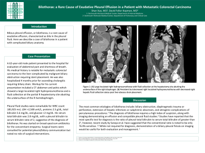
Figure: Figure 1: (A) Large loculated right hydropneumothorax with fluid collection at the hepatectomy site abutting the undersurface of the right diaphragm. (B) Persistent but decreased right loculated hydropneumothorax with decreased right hepatic fluid collection status post liver abscess drain placement.
Disclosures:
Shan Xue indicated no relevant financial relationships.
David Feller-Kopman indicated no relevant financial relationships.
Shan Xue, MD, David Feller-Kopman, MD. P0118 - Bilothorax: A Rare Cause of Exudative Pleural Effusion in a Patient with Metastatic Colorectal Carcinoma, ACG 2024 Annual Scientific Meeting Abstracts. Philadelphia, PA: American College of Gastroenterology.
