Tuesday Poster Session
Category: Esophagus
P3971 - Nothing by Mouth, Literally: Variceal Banding Complication
Tuesday, October 29, 2024
10:30 AM - 4:00 PM ET
Location: Exhibit Hall E

Has Audio

Frank Lee, BS
University of California Irvine School of Medicine
San Diego, CA
Presenting Author(s)
Frank Lee, BS1, Whitney Li, BA2, Erica Duh, MD3, Jason Samarasena, MD4, C Gregory Albers, MD4, Christina Ling, MD4
1University of California Irvine School of Medicine, San Diego, CA; 2University of California Irvine School of Medicine, San Jose, CA; 3University of California Irvine Digestive Health Institute, Orange, CA; 4University of California, Irvine, Orange, CA
Introduction: Variceal banding is used to prevent and treat esophageal variceal bleeding. Complete esophageal obstruction is an extremely rare but serious complication that can occur after esophageal variceal banding. Here, we present a case of complete esophageal obstruction after variceal banding.
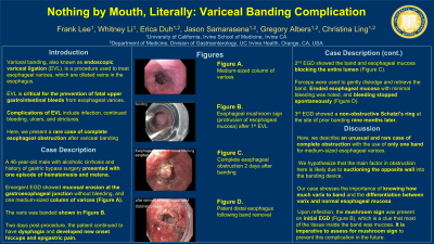
Case Description/Methods: A 46-year-old male with decompensated alcohol-induced cirrhosis and history of gastric bypass surgery (RYGB) presented to the emergency department with one episode of hematemesis and one episode of melena. He had stopped taking all medications one month prior, including his proton pump inhibitor, and had taken eight shots of espresso that day. Esophagogastroduodenoscopy (EGD) showed a small mucosal erosion at the gastroesophageal junction without active bleeding. One medium-sized column of esophageal varices was found, and one band was deployed for primary prophylaxis (Figure A).
One day later, the patient endorsed post-EGD dysphagia. Drinking small amounts of liquid led to nausea, neck pain, and non-bloody emesis. Two days post-procedure, dysphagia persisted along with new onset hiccups and epigastric pain. Abdominal X-ray was unremarkable. Repeat EGD showed the esophageal band surrounding blanched-appearing esophageal mucosa which blocked the entire lumen; small areas of sloughing in the distal esophagus were also found (Figure B). Rat tooth forceps were used to gently dislodge and retrieve the band. Circumferential eroded esophageal mucosa with minimal bleeding was noted, and bleeding stopped spontaneously (Figure C). Repeat EGD was recommended in one month to assess for possible dilation given anticipated scarring at the banding site. However, the patient did not return for repeat EGD until nine months later, which showed a non-obstructive Schatzi’s ring at the site of prior banding.
Discussion: Complete obstruction of the esophagus after endoscopic variceal banding is a very rare phenomenon. Fourteen other cases are reported in the literature. Most used multiple bands, with one case using up to eight. Here, we describe an unusual case of complete obstruction with the use of only one band for medium-sized esophageal varices. We hypothesize that the main factor in obstruction here is likely due to suctioning the opposite wall into the banding device. Upon reflection, the mushroom sign was present on initial EGD (Figure A), which is a clue that most of the tissue inside the band was mucosa. It is imperative to assess for mushroom sign to prevent this complication in the future.

Disclosures:
Frank Lee, BS1, Whitney Li, BA2, Erica Duh, MD3, Jason Samarasena, MD4, C Gregory Albers, MD4, Christina Ling, MD4. P3971 - Nothing by Mouth, Literally: Variceal Banding Complication, ACG 2024 Annual Scientific Meeting Abstracts. Philadelphia, PA: American College of Gastroenterology.
1University of California Irvine School of Medicine, San Diego, CA; 2University of California Irvine School of Medicine, San Jose, CA; 3University of California Irvine Digestive Health Institute, Orange, CA; 4University of California, Irvine, Orange, CA
Introduction: Variceal banding is used to prevent and treat esophageal variceal bleeding. Complete esophageal obstruction is an extremely rare but serious complication that can occur after esophageal variceal banding. Here, we present a case of complete esophageal obstruction after variceal banding.
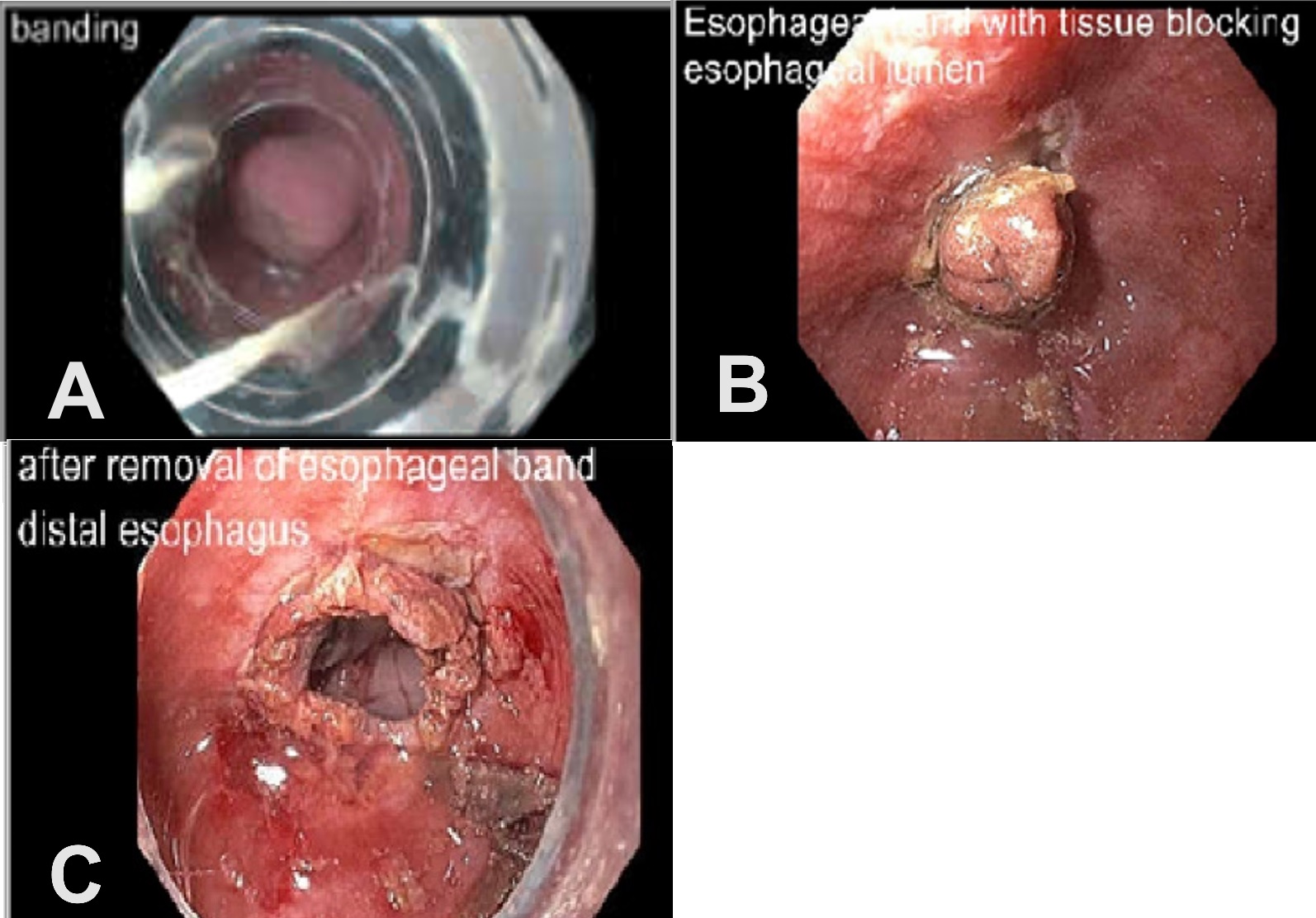
Case Description/Methods: A 46-year-old male with decompensated alcohol-induced cirrhosis and history of gastric bypass surgery (RYGB) presented to the emergency department with one episode of hematemesis and one episode of melena. He had stopped taking all medications one month prior, including his proton pump inhibitor, and had taken eight shots of espresso that day. Esophagogastroduodenoscopy (EGD) showed a small mucosal erosion at the gastroesophageal junction without active bleeding. One medium-sized column of esophageal varices was found, and one band was deployed for primary prophylaxis (Figure A).
One day later, the patient endorsed post-EGD dysphagia. Drinking small amounts of liquid led to nausea, neck pain, and non-bloody emesis. Two days post-procedure, dysphagia persisted along with new onset hiccups and epigastric pain. Abdominal X-ray was unremarkable. Repeat EGD showed the esophageal band surrounding blanched-appearing esophageal mucosa which blocked the entire lumen; small areas of sloughing in the distal esophagus were also found (Figure B). Rat tooth forceps were used to gently dislodge and retrieve the band. Circumferential eroded esophageal mucosa with minimal bleeding was noted, and bleeding stopped spontaneously (Figure C). Repeat EGD was recommended in one month to assess for possible dilation given anticipated scarring at the banding site. However, the patient did not return for repeat EGD until nine months later, which showed a non-obstructive Schatzi’s ring at the site of prior banding.
Discussion: Complete obstruction of the esophagus after endoscopic variceal banding is a very rare phenomenon. Fourteen other cases are reported in the literature. Most used multiple bands, with one case using up to eight. Here, we describe an unusual case of complete obstruction with the use of only one band for medium-sized esophageal varices. We hypothesize that the main factor in obstruction here is likely due to suctioning the opposite wall into the banding device. Upon reflection, the mushroom sign was present on initial EGD (Figure A), which is a clue that most of the tissue inside the band was mucosa. It is imperative to assess for mushroom sign to prevent this complication in the future.

Figure: Figure A: Esophageal mushroom sign after banding
Figure B: Esophageal obstruction after banding
Figure C: Removal of band relieving esophageal obstruction
Figure B: Esophageal obstruction after banding
Figure C: Removal of band relieving esophageal obstruction
Disclosures:
Frank Lee indicated no relevant financial relationships.
Whitney Li indicated no relevant financial relationships.
Erica Duh indicated no relevant financial relationships.
Jason Samarasena: Cook Medical – Consultant. Medtronic – Advisory Committee/Board Member, Consultant. Neptune Medical – Advisory Committee/Board Member, Consultant. Olympus – Advisory Committee/Board Member, Consultant. Ovesco – Advisory Committee/Board Member, Consultant, Speakers Bureau. SatisfAI – Stock-privately held company. Steris – Advisory Committee/Board Member.
C Gregory Albers: Nestle Health – Speakers Bureau. Phathom Pharmaceuticals – Speakers Bureau. Salix Pharmaceuticals – Speakers Bureau.
Christina Ling indicated no relevant financial relationships.
Frank Lee, BS1, Whitney Li, BA2, Erica Duh, MD3, Jason Samarasena, MD4, C Gregory Albers, MD4, Christina Ling, MD4. P3971 - Nothing by Mouth, Literally: Variceal Banding Complication, ACG 2024 Annual Scientific Meeting Abstracts. Philadelphia, PA: American College of Gastroenterology.
