Tuesday Poster Session
Category: Esophagus
P3974 - CMV Esophagitis Progressing to Severe Stricturing Disease Despite Treatment
Tuesday, October 29, 2024
10:30 AM - 4:00 PM ET
Location: Exhibit Hall E

Has Audio
- AV
Adam Vinall, MD
University of Texas Health Science Center
San Antonio, TX
Presenting Author(s)
Adam Vinall, MD1, Sylvia Keiser, DO2, Jawairia Memon, MD1, Juan Echavarria, MD3
1University of Texas Health Science Center, San Antonio, TX; 2UTHSCSA, San Antonio, TX; 3University of Texas Health San Antonio, San Antonio, TX
Introduction: Cytomegalovirus (CMV) esophagitis is a rare but significant complication typically observed in immunocompromised individuals, such as those with HIV/AIDS, organ transplant recipients, or patients undergoing immunosuppressive therapy. The most typical presentation is dysphagia, odynophagia, and weight loss. Initial response to treatment is typically good, though recurrence is common. Here we describe a case of CMV esophagitis progressing to severe esophageal stricture.
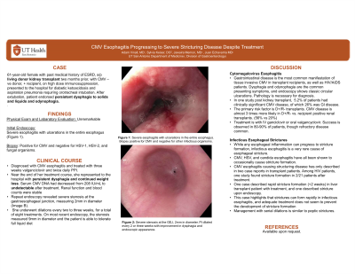
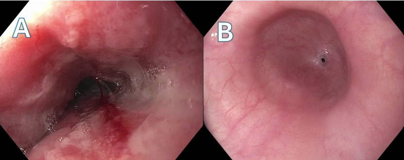
Case Description/Methods: A 61-year-old female with past medical history of end stage renal disease secondary to diabetes, status post living donor kidney transplant two months prior, with CMV negative donor and CMV positive recipient, on high dose immunosuppression, presented to the hospital for diabetic ketoacidosis and aspiration pneumonia requiring orotracheal intubation. After extubation, patient endorsed persistent dysphagia to solids and liquids and throat pain on swallowing. Speech therapy evaluation was consistent with severe pharyngeal dysphagia and she was kept N.P.O. Gastroenterology was consulted for percutaneous endoscopic gastrostomy (PEG) tube placement and she underwent upper endoscopy which revealed severe esophagitis with ulcerations in the entire esophagus (Image A). Biopsy was positive for CMV and negative for HSV-1, HSV-2, and fungal organisms. She was treated with three weeks of valganciclovir and twice daily omeprazole. Near the end of her treatment course, she represented to the hospital with persistent dysphagia and continued weight loss. Serum CMV DNA had decreased from 208 IU/mL to undetectable after treatment. Renal function and blood counts were stable. Repeat endoscopy revealed severe stenosis at the gastroesophageal junction, measuring 2mm in diameter (Image B). She underwent dilations every two to three weeks, for a total of eight treatments. On most recent endoscopy, the stenosis measured 9mm in diameter and the patient is able to tolerate full liquid diet.
Discussion: While benign esophageal strictures are common in reflux esophagitis, infectious esophagitis leading to stricturing disease is uncommon. Stricture formation in the context of CMV esophagitis is likely due to the chronic inflammatory response and subsequent fibrosis induced by persistent viral infection and ulceration. This case is unique due to the rapid progression from CMV esophagitis to a significant stricture within a short period, highlighting the aggressive nature of the disease despite treatment.

Disclosures:
Adam Vinall, MD1, Sylvia Keiser, DO2, Jawairia Memon, MD1, Juan Echavarria, MD3. P3974 - CMV Esophagitis Progressing to Severe Stricturing Disease Despite Treatment, ACG 2024 Annual Scientific Meeting Abstracts. Philadelphia, PA: American College of Gastroenterology.
1University of Texas Health Science Center, San Antonio, TX; 2UTHSCSA, San Antonio, TX; 3University of Texas Health San Antonio, San Antonio, TX
Introduction: Cytomegalovirus (CMV) esophagitis is a rare but significant complication typically observed in immunocompromised individuals, such as those with HIV/AIDS, organ transplant recipients, or patients undergoing immunosuppressive therapy. The most typical presentation is dysphagia, odynophagia, and weight loss. Initial response to treatment is typically good, though recurrence is common. Here we describe a case of CMV esophagitis progressing to severe esophageal stricture.
Case Description/Methods: A 61-year-old female with past medical history of end stage renal disease secondary to diabetes, status post living donor kidney transplant two months prior, with CMV negative donor and CMV positive recipient, on high dose immunosuppression, presented to the hospital for diabetic ketoacidosis and aspiration pneumonia requiring orotracheal intubation. After extubation, patient endorsed persistent dysphagia to solids and liquids and throat pain on swallowing. Speech therapy evaluation was consistent with severe pharyngeal dysphagia and she was kept N.P.O. Gastroenterology was consulted for percutaneous endoscopic gastrostomy (PEG) tube placement and she underwent upper endoscopy which revealed severe esophagitis with ulcerations in the entire esophagus (Image A). Biopsy was positive for CMV and negative for HSV-1, HSV-2, and fungal organisms. She was treated with three weeks of valganciclovir and twice daily omeprazole. Near the end of her treatment course, she represented to the hospital with persistent dysphagia and continued weight loss. Serum CMV DNA had decreased from 208 IU/mL to undetectable after treatment. Renal function and blood counts were stable. Repeat endoscopy revealed severe stenosis at the gastroesophageal junction, measuring 2mm in diameter (Image B). She underwent dilations every two to three weeks, for a total of eight treatments. On most recent endoscopy, the stenosis measured 9mm in diameter and the patient is able to tolerate full liquid diet.
Discussion: While benign esophageal strictures are common in reflux esophagitis, infectious esophagitis leading to stricturing disease is uncommon. Stricture formation in the context of CMV esophagitis is likely due to the chronic inflammatory response and subsequent fibrosis induced by persistent viral infection and ulceration. This case is unique due to the rapid progression from CMV esophagitis to a significant stricture within a short period, highlighting the aggressive nature of the disease despite treatment.

Figure: Image A. Diffuse moderate mucosal changes characterized by sloughing white mucosa were found in the entire esophagus. Biopsy was consistent with CMV esophagitis.
Image B. Follow up endoscopy after three weeks of treatment with valgancyclovir demonstrated improvement of the esophagitis but development of a severe esophageal stricture, 2mm in diameter.
Image B. Follow up endoscopy after three weeks of treatment with valgancyclovir demonstrated improvement of the esophagitis but development of a severe esophageal stricture, 2mm in diameter.
Disclosures:
Adam Vinall indicated no relevant financial relationships.
Sylvia Keiser indicated no relevant financial relationships.
Jawairia Memon indicated no relevant financial relationships.
Juan Echavarria indicated no relevant financial relationships.
Adam Vinall, MD1, Sylvia Keiser, DO2, Jawairia Memon, MD1, Juan Echavarria, MD3. P3974 - CMV Esophagitis Progressing to Severe Stricturing Disease Despite Treatment, ACG 2024 Annual Scientific Meeting Abstracts. Philadelphia, PA: American College of Gastroenterology.
