Tuesday Poster Session
Category: Esophagus
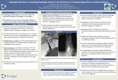
P4005 - Dysphagia Dilemmas: Unraveling Dysphagia Aortica in the Presence of a Cricopharyngeal Bar in an Elderly Woman
Tuesday, October 29, 2024
10:30 AM - 4:00 PM ET
Location: Exhibit Hall E

Has Audio

Landon Kozai, MD
University of Hawaii, John A. Burns School of Medicine
Honolulu, HI
Presenting Author(s)
Landon Kozai, MD1, Han Zhang, MD2
1University of Hawaii, John A. Burns School of Medicine, Honolulu, HI; 2Scripps Clinic, San Diego, CA
Introduction: Esophageal dysphagia is a common reason for gastroenterology consultation in the elderly. Here we describe a rare cause of dysphagia related to external compression of the esophagus by the aorta.
Case Description/Methods: A 94-year-old woman presented with two years of progressive dysphagia to solid foods, characterized by a sensation of food “piling up” in the lower chest. Her diet became limited to liquids, resulting in a loss of 8 lbs. She denied odynophagia, heartburn, and coughing or choking with meals. Her medical history was notable for a 3.7 cm distal thoracic aortic aneurysm, exaggerated thoracic kyphosis and several thoracolumbar compression fractures seen on CT imaging. The CT scan also noted tortuosity of her thoracic aorta.
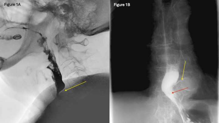
A modified barium swallow demonstrated normal oropharyngeal phase and the presence of a cricopharyngeal bar without any esophageal barium retention. On endoscopy, there was no evidence of strictures. The gastroesophageal junction appeared slightly spastic and due to prior patient request and her not tolerating high-resolution manometry, a trial of botox injection was performed to the distal esophagus. A 12 mm bougie dilation was also performed resulting in moderate mucosal disruption in the upper esophagus. She experienced no symptomatic improvement from the endoscopic therapy. Finally, a repeat barium esophagram was pursued, with the addition of a barium coated meal, which became retained in the distal esophagus due to the extrinsic compression from her tortuous distal thoracic aorta known as “dysphagia aortica.” Despite surgical consultation, the patient did not undergo surgery, and was managed conservatively with dietary modification.
Discussion: Occurring predominantly in elderly women, dysphagia aortica is associated with kyphoscoliosis and aortic dilation, aneurysm, or tortuosity. Radiographic evidence of external thoracic aortic compression of the esophagus against the crural diaphragm suggests the diagnosis. Dysphagia aortica is a diagnosis of exclusion; intraluminal obstruction and dysmotility should be ruled out. Treatment involves aortic endovascular repair, endoscopic stenting or dilation, feeding tube placement, or a modified oral diet.
This case demonstrates a diagnostic dilemma wherein patients can have multiple abnormal findings such as cricopharyngeal bar and thoracic aortic aneurysm. An esophagram with barium coated meal showed retention in the distal esophagus adjacent to a tortuous thoracic aorta, making it a useful tool to aid in diagnosis.

Disclosures:
Landon Kozai, MD1, Han Zhang, MD2. P4005 - Dysphagia Dilemmas: Unraveling Dysphagia Aortica in the Presence of a Cricopharyngeal Bar in an Elderly Woman, ACG 2024 Annual Scientific Meeting Abstracts. Philadelphia, PA: American College of Gastroenterology.
1University of Hawaii, John A. Burns School of Medicine, Honolulu, HI; 2Scripps Clinic, San Diego, CA
Introduction: Esophageal dysphagia is a common reason for gastroenterology consultation in the elderly. Here we describe a rare cause of dysphagia related to external compression of the esophagus by the aorta.
Case Description/Methods: A 94-year-old woman presented with two years of progressive dysphagia to solid foods, characterized by a sensation of food “piling up” in the lower chest. Her diet became limited to liquids, resulting in a loss of 8 lbs. She denied odynophagia, heartburn, and coughing or choking with meals. Her medical history was notable for a 3.7 cm distal thoracic aortic aneurysm, exaggerated thoracic kyphosis and several thoracolumbar compression fractures seen on CT imaging. The CT scan also noted tortuosity of her thoracic aorta.
A modified barium swallow demonstrated normal oropharyngeal phase and the presence of a cricopharyngeal bar without any esophageal barium retention. On endoscopy, there was no evidence of strictures. The gastroesophageal junction appeared slightly spastic and due to prior patient request and her not tolerating high-resolution manometry, a trial of botox injection was performed to the distal esophagus. A 12 mm bougie dilation was also performed resulting in moderate mucosal disruption in the upper esophagus. She experienced no symptomatic improvement from the endoscopic therapy. Finally, a repeat barium esophagram was pursued, with the addition of a barium coated meal, which became retained in the distal esophagus due to the extrinsic compression from her tortuous distal thoracic aorta known as “dysphagia aortica.” Despite surgical consultation, the patient did not undergo surgery, and was managed conservatively with dietary modification.
Discussion: Occurring predominantly in elderly women, dysphagia aortica is associated with kyphoscoliosis and aortic dilation, aneurysm, or tortuosity. Radiographic evidence of external thoracic aortic compression of the esophagus against the crural diaphragm suggests the diagnosis. Dysphagia aortica is a diagnosis of exclusion; intraluminal obstruction and dysmotility should be ruled out. Treatment involves aortic endovascular repair, endoscopic stenting or dilation, feeding tube placement, or a modified oral diet.
This case demonstrates a diagnostic dilemma wherein patients can have multiple abnormal findings such as cricopharyngeal bar and thoracic aortic aneurysm. An esophagram with barium coated meal showed retention in the distal esophagus adjacent to a tortuous thoracic aorta, making it a useful tool to aid in diagnosis.

Figure: Figure 1A: Cricopharyngeal bar (yellow arrow) demonstrated on modified barium swallow. Figure 1B: Esophagram with barium-coated scrambled eggs, retained in the distal esophagus (red arrow) proximal to the thoracic aorta (yellow arrow).
Disclosures:
Landon Kozai indicated no relevant financial relationships.
Han Zhang indicated no relevant financial relationships.
Landon Kozai, MD1, Han Zhang, MD2. P4005 - Dysphagia Dilemmas: Unraveling Dysphagia Aortica in the Presence of a Cricopharyngeal Bar in an Elderly Woman, ACG 2024 Annual Scientific Meeting Abstracts. Philadelphia, PA: American College of Gastroenterology.
