Tuesday Poster Session
Category: Esophagus
P4008 - An Unusual Presentation of HSV Esophagitis
Tuesday, October 29, 2024
10:30 AM - 4:00 PM ET
Location: Exhibit Hall E

- AS
Aditi Simlote, MD
Temple University Hospital
Philadelphia, PA
Presenting Author(s)
Aditi Simlote, MD, Neena Mohan, MD
Temple University Hospital, Philadelphia, PA
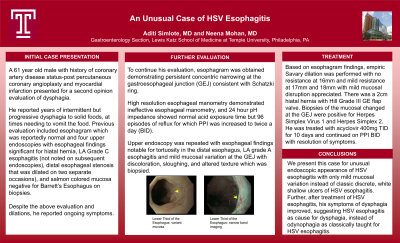
Introduction: A 61 year old male with history of coronary artery disease status-post percutaneous coronary angioplasty presented for a second opinion evaluation of dysphagia. He reported a few years of intermittent but progressive solid food dysphagia, at times needing to vomit the food. Previous evaluation included an esophagram showing a Schatzki ring and four upper endoscopies from 2021 to 2022. In 2021 EGDs noted a 1 cm hiatal hernia, esophageal stenosis s/p dilation, and LA Grade C esophagitis. In March 2022 EGD noted dilation of benign esophageal stenosis, and in June 2022 EGD did not show any stenosis, and biopsies of suspected Barrett’s esophagus were negative. Patient did not experience improvement of symptoms with dilations or twice daily PPI use.
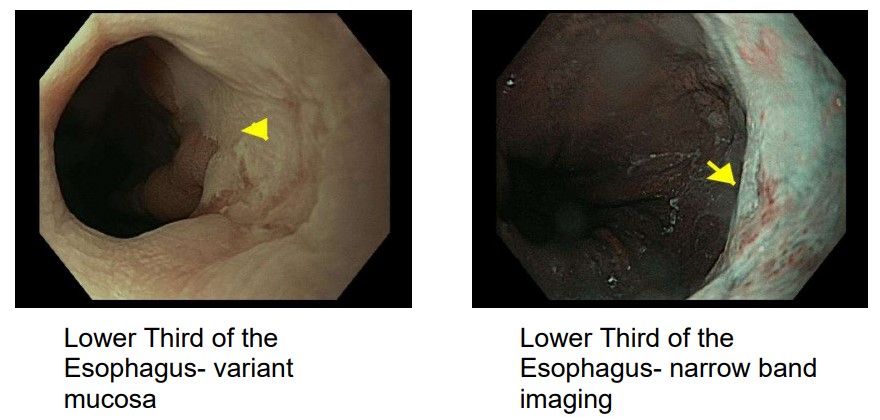
Case Description/Methods: He came to Temple hospital for evaluation November 2022. Esophagram demonstrated persistent concentric narrowing at the gastroesophageal junction (GEJ) consistent with Schatzki ring. High resolution esophageal manometry demonstrated ineffective esophageal manometry, and 24 hour pH impedance showed normal acid exposure time but 96 reflux episodes on once daily PPI. Upper endoscopy was repeated with esophageal findings notable for tortuosity in the distal esophagus, LA grade A esophagitis and mild mucosal variation at the GEJ with discoloration, sloughing, and altered texture which was biopsied. Based on esophagram findings, empiric Savary dilation was performed with no resistance at 16mm and mild resistance at 17mm and 18mm with mild mucosal disruption appreciated. There was a 2 cm hiatal hernia with Hill Grade III GE flap valve. Biopsies of the mucosal changes at the GEJ were positive for Herpes Simplex Virus 1 and Herpes Simplex Virus 2. He was treated with acyclovir 400mg TID for 10 days, with resolution of dysphagia symptoms.
Discussion: We present this case for the unusual endoscopic appearance of HSV esophagitis with only mild mucosal variation instead of classic discrete, shallow ulcers and for the chronicity of dysphagia rather than acute symptoms. Additionally, HSV-2 is a rare cause of HSV esophagitis in immunocompetent patients. This case reaffirms the importance of a careful esophageal examination to identify subtle abnormal findings.

Disclosures:
Aditi Simlote, MD, Neena Mohan, MD. P4008 - An Unusual Presentation of HSV Esophagitis, ACG 2024 Annual Scientific Meeting Abstracts. Philadelphia, PA: American College of Gastroenterology.
Temple University Hospital, Philadelphia, PA
Introduction: A 61 year old male with history of coronary artery disease status-post percutaneous coronary angioplasty presented for a second opinion evaluation of dysphagia. He reported a few years of intermittent but progressive solid food dysphagia, at times needing to vomit the food. Previous evaluation included an esophagram showing a Schatzki ring and four upper endoscopies from 2021 to 2022. In 2021 EGDs noted a 1 cm hiatal hernia, esophageal stenosis s/p dilation, and LA Grade C esophagitis. In March 2022 EGD noted dilation of benign esophageal stenosis, and in June 2022 EGD did not show any stenosis, and biopsies of suspected Barrett’s esophagus were negative. Patient did not experience improvement of symptoms with dilations or twice daily PPI use.
Case Description/Methods: He came to Temple hospital for evaluation November 2022. Esophagram demonstrated persistent concentric narrowing at the gastroesophageal junction (GEJ) consistent with Schatzki ring. High resolution esophageal manometry demonstrated ineffective esophageal manometry, and 24 hour pH impedance showed normal acid exposure time but 96 reflux episodes on once daily PPI. Upper endoscopy was repeated with esophageal findings notable for tortuosity in the distal esophagus, LA grade A esophagitis and mild mucosal variation at the GEJ with discoloration, sloughing, and altered texture which was biopsied. Based on esophagram findings, empiric Savary dilation was performed with no resistance at 16mm and mild resistance at 17mm and 18mm with mild mucosal disruption appreciated. There was a 2 cm hiatal hernia with Hill Grade III GE flap valve. Biopsies of the mucosal changes at the GEJ were positive for Herpes Simplex Virus 1 and Herpes Simplex Virus 2. He was treated with acyclovir 400mg TID for 10 days, with resolution of dysphagia symptoms.
Discussion: We present this case for the unusual endoscopic appearance of HSV esophagitis with only mild mucosal variation instead of classic discrete, shallow ulcers and for the chronicity of dysphagia rather than acute symptoms. Additionally, HSV-2 is a rare cause of HSV esophagitis in immunocompetent patients. This case reaffirms the importance of a careful esophageal examination to identify subtle abnormal findings.

Figure: Endoscopic appearance of mucosal variation under white light and narrow band imaging.
Disclosures:
Aditi Simlote indicated no relevant financial relationships.
Neena Mohan indicated no relevant financial relationships.
Aditi Simlote, MD, Neena Mohan, MD. P4008 - An Unusual Presentation of HSV Esophagitis, ACG 2024 Annual Scientific Meeting Abstracts. Philadelphia, PA: American College of Gastroenterology.
