Tuesday Poster Session
Category: Esophagus
P4017 - Primary Esophageal Malignant Melanoma: A Rare Endoscopic Finding
Tuesday, October 29, 2024
10:30 AM - 4:00 PM ET
Location: Exhibit Hall E

Has Audio

Nikitha Chellapuram, MD
Centinela Hospital Medical Center
Los Angeles, CA
Presenting Author(s)
Yamini Katamreddy, MD1, Nikitha Chellapuram, MD2, Saikiran Mandyam, MD3, Sri Laxmi Valasareddi, MD3
1West Anaheim Medical Center, Anaheim, CA; 2Centinela Hospital Medical Center, Los Angeles, CA; 3Southeast Health, Dothan, AL
Introduction: Primary esophageal melanoma is a rare entity, accounting for 0.1-0.2% of all esophageal malignancies. The prognosis is poor, with a 5-year survival rate of less than 5%. We report on a case of primary malignant esophageal melanoma.
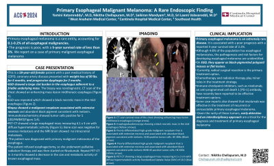
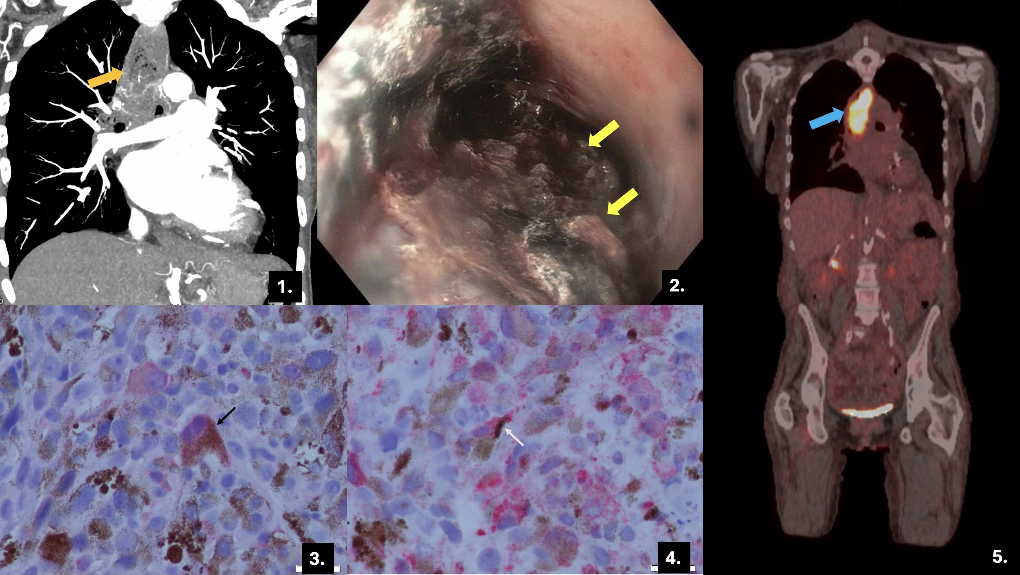
Case Description/Methods: This is a 58-year-old female patient with a past medical history of COPD, coronary artery disease presented with weight loss of 90 lbs. for 6 months, and progressive dysphagia for 2 months. Esophagoduodenoscopy showed a large clot burden in the esophagus adherent to a friable underlying mass. The biopsy was nondiagnostic. CT scan of the chest showed an enhancing mass lesion midthoracic esophagus (Figure 1). EGD was repeated which showed a black necrotic mass in the mid-esophagus (Figure 2). Biopsy showed a malignant neoplasm associated with extensive necrosis and abundant black pigment consistent with melanin. Immunohistochemistry showed tumor cells positive for S 100/HMB-45(Figure 3,4). PET-CT showed a large esophageal mass measuring 4.1 x 3 cm with diffuse hypermetabolic activity (Figure 5). Bone scan was negative for osseous metastasis and the MRI brain showed no intracranial metastasis. The patient was diagnosed with primary malignant melanoma of the esophagus. The patient refused esophagectomy, so she underwent palliative radiation therapy and was then started on Nivolumab. Repeat PET-CT in 3 months showed a decrease in the size and metabolic activity of known esophageal mass.
Discussion: Primary esophageal melanoma is an extremely rare disease. It is associated with a poor prognosis with a reported 5-year survival rate of 2.2%. Although 4-8% of the population has esophageal melanocytes, the pathogenesis and risk factors for developing esophageal melanoma are unidentified. On EGD, they appear as black-pigmented polypoid masses or flat lesions. Currently, radical surgical resection is the primary treatment option. Chemotherapy and radiation therapy play minor roles in the treatment strategy. Immune checkpoint inhibitors, such as nivolumab, an anti-programmed cell death 1 [PD-1] antibody, have recently been reported to be effective treatment options. Some case reports also showed that nivolumab was effective in the treatment of recurrent or unresectable primary esophageal melanoma. Given the rarity of these tumors, early detection and an interdisciplinary approach are critical for the diagnosis and treatment of primary esophageal melanoma.

Disclosures:
Yamini Katamreddy, MD1, Nikitha Chellapuram, MD2, Saikiran Mandyam, MD3, Sri Laxmi Valasareddi, MD3. P4017 - Primary Esophageal Malignant Melanoma: A Rare Endoscopic Finding, ACG 2024 Annual Scientific Meeting Abstracts. Philadelphia, PA: American College of Gastroenterology.
1West Anaheim Medical Center, Anaheim, CA; 2Centinela Hospital Medical Center, Los Angeles, CA; 3Southeast Health, Dothan, AL
Introduction: Primary esophageal melanoma is a rare entity, accounting for 0.1-0.2% of all esophageal malignancies. The prognosis is poor, with a 5-year survival rate of less than 5%. We report on a case of primary malignant esophageal melanoma.
Case Description/Methods: This is a 58-year-old female patient with a past medical history of COPD, coronary artery disease presented with weight loss of 90 lbs. for 6 months, and progressive dysphagia for 2 months. Esophagoduodenoscopy showed a large clot burden in the esophagus adherent to a friable underlying mass. The biopsy was nondiagnostic. CT scan of the chest showed an enhancing mass lesion midthoracic esophagus (Figure 1). EGD was repeated which showed a black necrotic mass in the mid-esophagus (Figure 2). Biopsy showed a malignant neoplasm associated with extensive necrosis and abundant black pigment consistent with melanin. Immunohistochemistry showed tumor cells positive for S 100/HMB-45(Figure 3,4). PET-CT showed a large esophageal mass measuring 4.1 x 3 cm with diffuse hypermetabolic activity (Figure 5). Bone scan was negative for osseous metastasis and the MRI brain showed no intracranial metastasis. The patient was diagnosed with primary malignant melanoma of the esophagus. The patient refused esophagectomy, so she underwent palliative radiation therapy and was then started on Nivolumab. Repeat PET-CT in 3 months showed a decrease in the size and metabolic activity of known esophageal mass.
Discussion: Primary esophageal melanoma is an extremely rare disease. It is associated with a poor prognosis with a reported 5-year survival rate of 2.2%. Although 4-8% of the population has esophageal melanocytes, the pathogenesis and risk factors for developing esophageal melanoma are unidentified. On EGD, they appear as black-pigmented polypoid masses or flat lesions. Currently, radical surgical resection is the primary treatment option. Chemotherapy and radiation therapy play minor roles in the treatment strategy. Immune checkpoint inhibitors, such as nivolumab, an anti-programmed cell death 1 [PD-1] antibody, have recently been reported to be effective treatment options. Some case reports also showed that nivolumab was effective in the treatment of recurrent or unresectable primary esophageal melanoma. Given the rarity of these tumors, early detection and an interdisciplinary approach are critical for the diagnosis and treatment of primary esophageal melanoma.

Figure: Figure 1: CT scan coronal view of the chest showing enhancing mass lesion midthoracic esophagus (orange arrow).
Figure 2: Esophagoduodenoscopy showing a black necrotic mass in the mid-esophagus (Yellow arrows).
Figure 3: Poorly differentiated high-grade malignant neoplasm that is associated with extensive necrosis and associated with abundant black pigment consistent with melanin. S100 positive tumor cells. HE 400x (Black arrow).
Figure 4: Poorly differentiated high-grade malignant neoplasm that is associated with extensive necrosis and associated with abundant black pigment consistent with melanin
HMB-45 positive tumor cells. HE 400x (White arrow).
Figure 5: PET-CT showing a large esophageal mass measuring 4.1 x 3 cm with diffuse hypermetabolic activity Standardized Uptake Value (SUV) of 18.3 (blue arrow).
Disclosures:
Yamini Katamreddy indicated no relevant financial relationships.
Nikitha Chellapuram indicated no relevant financial relationships.
Saikiran Mandyam indicated no relevant financial relationships.
Sri Laxmi Valasareddi indicated no relevant financial relationships.
Yamini Katamreddy, MD1, Nikitha Chellapuram, MD2, Saikiran Mandyam, MD3, Sri Laxmi Valasareddi, MD3. P4017 - Primary Esophageal Malignant Melanoma: A Rare Endoscopic Finding, ACG 2024 Annual Scientific Meeting Abstracts. Philadelphia, PA: American College of Gastroenterology.
