Tuesday Poster Session
Category: Esophagus
P4022 - Double Barrel Esophagus in a Patient with Longstanding Retained Foreign Body
Tuesday, October 29, 2024
10:30 AM - 4:00 PM ET
Location: Exhibit Hall E

Has Audio

Lila Bilsky, BS
McGovern Medical School at UTHealth
Houston, TX
Presenting Author(s)
Lila Bilsky, BS, Christine Catinis, MD, MS
McGovern Medical School at UTHealth, Houston, TX
Introduction: Foreign body (FB) ingestion occurs frequently in children, with coins being the most common culprit. While the majority of ingested FBs pass spontaneously, complications are more likely to occur when objects are retained for greater than 24 hours. Retained FBs can result in various complications, including tracheal compression and mucosal erosion with migration to nearby structures. Here, we report a case of a long-standing ingested foreign body that led to double barrel esophagus and FB material in the trachea.
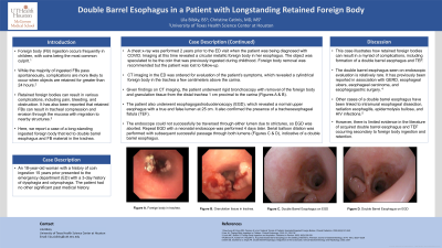
Case Description/Methods: An 18-year-old female with a history of coin ingestion 15 years prior presented to the emergency department with a 3 day history of dysphagia and odynophagia. CT imaging revealed a cylindrical foreign body in the trachea a few centimeters above the carina. Of note, she had a chest x-ray performed 2 years ago when being diagnosed with COVID which revealed a circular metallic foreign body in her esophagus. The object was speculated to be the previously ingested coin so foreign body removal was recommended, but the patient was lost to follow-up. Given her CT findings, she underwent rigid bronchoscopy with removal of the foreign body and granulation tissue from the distal trachea 1 cm proximal to the carina. She also underwent esophagogastroduodenoscopy (EGD), which revealed a normal upper esophagus with a true and false lumen at 25 cm. It also confirmed presence of a tracheoesophageal fistula (TEF). The endoscope could not successfully be traversed through either lumen due to strictures, so EGD was aborted. Repeat EGD with a neonatal endoscope was performed 4 days later. Serial balloon dilation was performed with subsequent successful passage through both lumens, indicative of double barrel esophagus.
Discussion: This case illustrates how retained foreign bodies can result in a myriad of complications, including formation of a double barrel esophagus and TEF. The double barrel esophagus seen on endoscopic evaluation is relatively rare and has previously been reported in association with GERD, esophageal ulcers, esophageal carcinoma, and esophagogastric surgery. Other cases have been linked to intramural esophageal dissection, radiation esophagitis, epidermolysis bullosa, and HIV infections. However, there is limited evidence in the literature of acquired double barrel esophagus occurring secondary to foreign body ingestion and retention.

Disclosures:
Lila Bilsky, BS, Christine Catinis, MD, MS. P4022 - Double Barrel Esophagus in a Patient with Longstanding Retained Foreign Body, ACG 2024 Annual Scientific Meeting Abstracts. Philadelphia, PA: American College of Gastroenterology.
McGovern Medical School at UTHealth, Houston, TX
Introduction: Foreign body (FB) ingestion occurs frequently in children, with coins being the most common culprit. While the majority of ingested FBs pass spontaneously, complications are more likely to occur when objects are retained for greater than 24 hours. Retained FBs can result in various complications, including tracheal compression and mucosal erosion with migration to nearby structures. Here, we report a case of a long-standing ingested foreign body that led to double barrel esophagus and FB material in the trachea.
Case Description/Methods: An 18-year-old female with a history of coin ingestion 15 years prior presented to the emergency department with a 3 day history of dysphagia and odynophagia. CT imaging revealed a cylindrical foreign body in the trachea a few centimeters above the carina. Of note, she had a chest x-ray performed 2 years ago when being diagnosed with COVID which revealed a circular metallic foreign body in her esophagus. The object was speculated to be the previously ingested coin so foreign body removal was recommended, but the patient was lost to follow-up. Given her CT findings, she underwent rigid bronchoscopy with removal of the foreign body and granulation tissue from the distal trachea 1 cm proximal to the carina. She also underwent esophagogastroduodenoscopy (EGD), which revealed a normal upper esophagus with a true and false lumen at 25 cm. It also confirmed presence of a tracheoesophageal fistula (TEF). The endoscope could not successfully be traversed through either lumen due to strictures, so EGD was aborted. Repeat EGD with a neonatal endoscope was performed 4 days later. Serial balloon dilation was performed with subsequent successful passage through both lumens, indicative of double barrel esophagus.
Discussion: This case illustrates how retained foreign bodies can result in a myriad of complications, including formation of a double barrel esophagus and TEF. The double barrel esophagus seen on endoscopic evaluation is relatively rare and has previously been reported in association with GERD, esophageal ulcers, esophageal carcinoma, and esophagogastric surgery. Other cases have been linked to intramural esophageal dissection, radiation esophagitis, epidermolysis bullosa, and HIV infections. However, there is limited evidence in the literature of acquired double barrel esophagus occurring secondary to foreign body ingestion and retention.

Figure: Image A shows the presence of a foreign body in the trachea. Image B displays granulation tissue in the trachea. Images C and D show double barrel esophagus on endoscopy.
Disclosures:
Lila Bilsky indicated no relevant financial relationships.
Christine Catinis indicated no relevant financial relationships.
Lila Bilsky, BS, Christine Catinis, MD, MS. P4022 - Double Barrel Esophagus in a Patient with Longstanding Retained Foreign Body, ACG 2024 Annual Scientific Meeting Abstracts. Philadelphia, PA: American College of Gastroenterology.
