Tuesday Poster Session
Category: Esophagus
P4036 - Behind the Swallow: Unraveling Dysphagia Lusoria with Esophageal Manometry
Tuesday, October 29, 2024
10:30 AM - 4:00 PM ET
Location: Exhibit Hall E

Has Audio
- DJ
Danielle Jackson, DO
UT Health Science Center
Tyler, TX
Presenting Author(s)
Danielle Jackson, DO1, Layth Alzubaidy, MD2, Bolarinwa Olusola, MD3
1UT Health Science Center, Tyler, TX; 2University of Texas Health Science Center, Tyler, TX; 3University of Texas Health East Texas Physicians, Tyler, TX
Introduction: Dysphagia lusoria (DL) is a rare cause of esophageal dysphagia. it is characterized by an external force compressing the esophagus which is due to an aberrant right subclavian artery (ARSA) that is present at birth and can present later in life as an obscure cause of dysphagia.
Case Description/Methods: We present a case of a 63-year-old African American female who presented with dysphagia in the clinic for many years. Laboratory workup and physical exam were rather unremarkable. Upper endoscopy was pursued which revealed a normal-appearing esophageal mucosa and gastroesophageal junction. However, there was a prominence of the upper esophagus indicating a possible extrinsic nature of dysphagia (Figure 1A). Random esophageal biopsies were negative for eosinophilic esophagitis. A modified barium swallow demonstrated slight narrowing of the left side of the esophagus at the level of the aortic arch (Figure 1B). High-resolution esophageal manometry (HRM) was pursued. The HRM Chicago 4.0 protocol was followed and revealed no evidence of disorders of peristalsis. However, a closer look at the HRM pictorial representation showed a pulsatile area of pressure in the proximal esophagus (Figure 1C). CT scan of the chest demonstrated a normal esophagus with post-surgical findings of the carotid subclavian bypass, and calcified origin of the aberrant right subclavian artery. These findings confirm the diagnosis of DL.
Discussion: This case underscores the importance of expanding the differential diagnosis when evaluating patients with dysphagia lusoria. Initial evaluation typically involves endoscopy, which reveals significant findings in only about half of the cases. Currently, barium swallow is the preferred diagnostic tool, as it can assess extrinsic compression of the esophagus, as demonstrated in this case. Treatment varies based on symptom severity and can range from conservative lifestyle modifications to surgical vascular repair. Recent studies indicate an association between high-resolution manometry (HRM) and pulsatile pressure zones (PPZ) in older populations. Although the role of HRM in diagnosing DL remains uncertain, recognizing these pressure zones on HRM in the appropriate clinical context—considering age and medical history—may prompt further investigation, leading to the diagnosis of such an uncommon etiology of dysphagia.

Disclosures:
Danielle Jackson, DO1, Layth Alzubaidy, MD2, Bolarinwa Olusola, MD3. P4036 - Behind the Swallow: Unraveling Dysphagia Lusoria with Esophageal Manometry, ACG 2024 Annual Scientific Meeting Abstracts. Philadelphia, PA: American College of Gastroenterology.
1UT Health Science Center, Tyler, TX; 2University of Texas Health Science Center, Tyler, TX; 3University of Texas Health East Texas Physicians, Tyler, TX
Introduction: Dysphagia lusoria (DL) is a rare cause of esophageal dysphagia. it is characterized by an external force compressing the esophagus which is due to an aberrant right subclavian artery (ARSA) that is present at birth and can present later in life as an obscure cause of dysphagia.
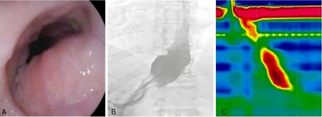
Case Description/Methods: We present a case of a 63-year-old African American female who presented with dysphagia in the clinic for many years. Laboratory workup and physical exam were rather unremarkable. Upper endoscopy was pursued which revealed a normal-appearing esophageal mucosa and gastroesophageal junction. However, there was a prominence of the upper esophagus indicating a possible extrinsic nature of dysphagia (Figure 1A). Random esophageal biopsies were negative for eosinophilic esophagitis. A modified barium swallow demonstrated slight narrowing of the left side of the esophagus at the level of the aortic arch (Figure 1B). High-resolution esophageal manometry (HRM) was pursued. The HRM Chicago 4.0 protocol was followed and revealed no evidence of disorders of peristalsis. However, a closer look at the HRM pictorial representation showed a pulsatile area of pressure in the proximal esophagus (Figure 1C). CT scan of the chest demonstrated a normal esophagus with post-surgical findings of the carotid subclavian bypass, and calcified origin of the aberrant right subclavian artery. These findings confirm the diagnosis of DL.
Discussion: This case underscores the importance of expanding the differential diagnosis when evaluating patients with dysphagia lusoria. Initial evaluation typically involves endoscopy, which reveals significant findings in only about half of the cases. Currently, barium swallow is the preferred diagnostic tool, as it can assess extrinsic compression of the esophagus, as demonstrated in this case. Treatment varies based on symptom severity and can range from conservative lifestyle modifications to surgical vascular repair. Recent studies indicate an association between high-resolution manometry (HRM) and pulsatile pressure zones (PPZ) in older populations. Although the role of HRM in diagnosing DL remains uncertain, recognizing these pressure zones on HRM in the appropriate clinical context—considering age and medical history—may prompt further investigation, leading to the diagnosis of such an uncommon etiology of dysphagia.

Figure: Figure 1: (A) endoscopic image showing esophageal with likely prominent aortic arch. (B) modified barium swallow demonstrating slight narrowing of the left side of the esophagus at the level of the aortic arch. (C) high-resolution esophageal manometry with pulsatile pressures noted at the dotted yellow line.
Disclosures:
Danielle Jackson indicated no relevant financial relationships.
Layth Alzubaidy indicated no relevant financial relationships.
Bolarinwa Olusola indicated no relevant financial relationships.
Danielle Jackson, DO1, Layth Alzubaidy, MD2, Bolarinwa Olusola, MD3. P4036 - Behind the Swallow: Unraveling Dysphagia Lusoria with Esophageal Manometry, ACG 2024 Annual Scientific Meeting Abstracts. Philadelphia, PA: American College of Gastroenterology.
