Tuesday Poster Session
Category: GI Bleeding
P4193 - Gastric and Colonic Leiomyosarcoma: An Extremely Rare Cause of Occult GI Bleed
Tuesday, October 29, 2024
10:30 AM - 4:00 PM ET
Location: Exhibit Hall E


Ibrar Atiq, MD
United Health Services, Wilson Medical Center
Binghamton, NY
Presenting Author(s)
Ibrar Atiq, MD1, Khadija Raza, MD1, Gowthami Ramar, MD2, Yasir Ahmed, MD1, Mohammad Abidi, MD1, Toseef Javaid, MD1
1United Health Services, Wilson Medical Center, Binghamton, NY; 2United Health Services, Binghamton, NY
Introduction: Leiomyosarcoma (LMS) is an extremely rare and different from Gastrointestinal stromal tumor (GIST) accounting for < 1% of gastrointestinal (GI) tumors. Here we report a case of this aggressive tumor presented as occult GI bleed.
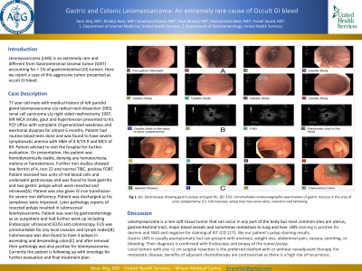
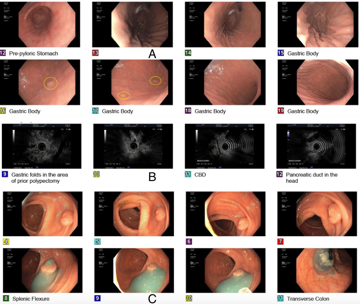
Case Description/Methods: 77 year old male with medical history of left parotid gland leiomyosarcoma s/p radical neck dissection 2003, renal cell carcinoma s/p right sided nephrectomy 1997, left MCA stroke, gout and hypertension presented to his PCP office with complaint of generalized weakness and exertional dyspnea for almost 6 months. Patient had routine blood tests done and was found to have severe symptomatic anemia with H&H of 4.9/19.9 and MCV of 69. Patient advised to visit the hospital for further evaluation. On presentation, the patient was hemodynamically stable, denying any hematochezia, melena or hematemesis. Further iron studies showed low ferritin of 4, Iron 22 and normal TIBC, positive FOBT. Patient received two units of red blood cells and underwent gastroscopy and was found to have gastritis and two gastric polyps which were resected and retrieved[A]. Patient was also given IV iron transfusion for severe iron deficiency. Patient was discharged as his symptoms were improved. Later pathology reports of resected polyps resulted in submucosal leiomyosarcoma. Patient was seen by gastroenterology as an outpatient and had further work up including Endoscopic ultrasound (EUS) and colonoscopy. EUS was unremarkable for any local invasion and lymph nodes[B]. Colonoscopy was also found to have 3 polyps in ascending and descending colon[C] and after removal their pathology was also positive for leiomyosarcoma. Currently the patient is following up with oncology for further evaluation and final treatment plan.
Discussion: Leiomyosarcoma is a rare soft tissue tumor that can occur in any part of the body but most common sites are uterus, gastrointestinal tract, major blood vessels and sometimes metastasis to lung and liver. LMS staining is positive for desmin and SMA and negative for staining of KIT (CD 117), like our patient’s polyp staining results.
Gastric LMS is usually asymptomatic but can present with anorexia, weight loss, abdominal pain, nausea, vomiting, or bleeding. Their diagnosis is confirmed with Endoscopy and biopsy of the tumor/polyp.
Local tumors with size >2 cm surgical resection is the preferred method with or without neoadjuvant therapy. For metastatic disease, benefits of adjuvant chemotherapy are controversial as there is a high risk of recurrence.

Disclosures:
Ibrar Atiq, MD1, Khadija Raza, MD1, Gowthami Ramar, MD2, Yasir Ahmed, MD1, Mohammad Abidi, MD1, Toseef Javaid, MD1. P4193 - Gastric and Colonic Leiomyosarcoma: An Extremely Rare Cause of Occult GI Bleed, ACG 2024 Annual Scientific Meeting Abstracts. Philadelphia, PA: American College of Gastroenterology.
1United Health Services, Wilson Medical Center, Binghamton, NY; 2United Health Services, Binghamton, NY
Introduction: Leiomyosarcoma (LMS) is an extremely rare and different from Gastrointestinal stromal tumor (GIST) accounting for < 1% of gastrointestinal (GI) tumors. Here we report a case of this aggressive tumor presented as occult GI bleed.
Case Description/Methods: 77 year old male with medical history of left parotid gland leiomyosarcoma s/p radical neck dissection 2003, renal cell carcinoma s/p right sided nephrectomy 1997, left MCA stroke, gout and hypertension presented to his PCP office with complaint of generalized weakness and exertional dyspnea for almost 6 months. Patient had routine blood tests done and was found to have severe symptomatic anemia with H&H of 4.9/19.9 and MCV of 69. Patient advised to visit the hospital for further evaluation. On presentation, the patient was hemodynamically stable, denying any hematochezia, melena or hematemesis. Further iron studies showed low ferritin of 4, Iron 22 and normal TIBC, positive FOBT. Patient received two units of red blood cells and underwent gastroscopy and was found to have gastritis and two gastric polyps which were resected and retrieved[A]. Patient was also given IV iron transfusion for severe iron deficiency. Patient was discharged as his symptoms were improved. Later pathology reports of resected polyps resulted in submucosal leiomyosarcoma. Patient was seen by gastroenterology as an outpatient and had further work up including Endoscopic ultrasound (EUS) and colonoscopy. EUS was unremarkable for any local invasion and lymph nodes[B]. Colonoscopy was also found to have 3 polyps in ascending and descending colon[C] and after removal their pathology was also positive for leiomyosarcoma. Currently the patient is following up with oncology for further evaluation and final treatment plan.
Discussion: Leiomyosarcoma is a rare soft tissue tumor that can occur in any part of the body but most common sites are uterus, gastrointestinal tract, major blood vessels and sometimes metastasis to lung and liver. LMS staining is positive for desmin and SMA and negative for staining of KIT (CD 117), like our patient’s polyp staining results.
Gastric LMS is usually asymptomatic but can present with anorexia, weight loss, abdominal pain, nausea, vomiting, or bleeding. Their diagnosis is confirmed with Endoscopy and biopsy of the tumor/polyp.
Local tumors with size >2 cm surgical resection is the preferred method with or without neoadjuvant therapy. For metastatic disease, benefits of adjuvant chemotherapy are controversial as there is a high risk of recurrence.

Figure: A. Gastroscopy showing gastric polyps and gastritis. B. EUS: Unremarkable endosonographic examination of gastric
mucosa in the area of prior polypectomy. C. Colonoscopy: polyp near ileo-cecal valve, resection and tattooing.
mucosa in the area of prior polypectomy. C. Colonoscopy: polyp near ileo-cecal valve, resection and tattooing.
Disclosures:
Ibrar Atiq indicated no relevant financial relationships.
Khadija Raza indicated no relevant financial relationships.
Gowthami Ramar indicated no relevant financial relationships.
Yasir Ahmed indicated no relevant financial relationships.
Mohammad Abidi indicated no relevant financial relationships.
Toseef Javaid indicated no relevant financial relationships.
Ibrar Atiq, MD1, Khadija Raza, MD1, Gowthami Ramar, MD2, Yasir Ahmed, MD1, Mohammad Abidi, MD1, Toseef Javaid, MD1. P4193 - Gastric and Colonic Leiomyosarcoma: An Extremely Rare Cause of Occult GI Bleed, ACG 2024 Annual Scientific Meeting Abstracts. Philadelphia, PA: American College of Gastroenterology.
