Tuesday Poster Session
Category: GI Bleeding
P4222 - A Rare Case of Ileal Neuroendocrine Tumor Presenting With Gastrointestinal Bleeding
Tuesday, October 29, 2024
10:30 AM - 4:00 PM ET
Location: Exhibit Hall E

Has Audio

Cindy Traboulsi, MD
University of Pittsburgh Medical Center
Pittsburgh, PA
Presenting Author(s)
Cindy Traboulsi, MD, Andrew Groff, MD, Gabriel Marrero-Rivera, MD, Kenneth Fasanella, MD
University of Pittsburgh Medical Center, Pittsburgh, PA
Introduction: Small bowel neuroendocrine tumors (NET) are rare and mostly located within 60cm of the ileocecal valve.1 Patients with ileal NET are typically asymptomatic but may present with abdominal pain and more rarely, gastrointestinal (GI) bleeding. We present a rare case of ileal NET in a patient with hemophilia B presenting as acute lower GI bleeding.
Case Description/Methods: A 55-year-old man with a history of hemophilia B, diverticulosis, and a prior episode of GI bleeding without a known source presented with a one-day history of 10 liquid, maroon-colored bowel movements. On arrival, he was tachycardic but normotensive. His hemoglobin (Hgb) was 12.2g/dL with an unclear baseline. While in the ED, he had two episodes of syncope associated with bright red blood per rectum. He received an infusion of factor IX and was sent for CT Angiography (CTA). There was evidence of blood in the distal ileum without active extravasation and no embolization was performed. Despite factor IX replacement, he continued to have maroon-colored bowel movements and his Hgb dropped to 7.5g/dL. Given ongoing bleeding without signs of active extravasation, a colonoscopy was performed and found multiple small-mouthed non-bleeding diverticula. The distal ileum contained a 1cm submucosal noncircumferential non-obstructing medium-sized mass with a 5mm clean-based ulceration, suspicious for an ileal NET about 50cm proximal to the ileocecal valve (Figure). No bleeding was present. Biopsies were taken for histology. GI Surgical Oncology was consulted and performed a small bowel resection with lymph node dissection. Pathology was consistent with a well-differentiated NET (WHO G1) involving the small intestinal mucosa with evidence of metastatic NET in 7 out of 8 lymph nodes. The tumor cells were positive for synaptophysin and INSM1 with a KI67 index of < 1%. Post-op, he did not have further bleeding and his Hgb stabilized.
Discussion: We present a rare case of a patient with hemophilia B who developed acute lower GI bleeding with evidence of recent ileal bleeding on CTA and was found to have a well-differentiated NET. He then underwent surgical resection which is crucial in the management of ileal NETs as the rate of metastatic disease is ~50%.1 A high index of suspicion for ileal NETs is required in patients with signs of ileal bleeding on CTA for a timely diagnosis. This case reinforces the importance of endoscopic evaluation in patients with concerns for ileal bleeding.
1Ahmed, M. World J Gastrointest Oncol. 2020;12(8):791-807.

Disclosures:
Cindy Traboulsi, MD, Andrew Groff, MD, Gabriel Marrero-Rivera, MD, Kenneth Fasanella, MD. P4222 - A Rare Case of Ileal Neuroendocrine Tumor Presenting With Gastrointestinal Bleeding, ACG 2024 Annual Scientific Meeting Abstracts. Philadelphia, PA: American College of Gastroenterology.
University of Pittsburgh Medical Center, Pittsburgh, PA
Introduction: Small bowel neuroendocrine tumors (NET) are rare and mostly located within 60cm of the ileocecal valve.1 Patients with ileal NET are typically asymptomatic but may present with abdominal pain and more rarely, gastrointestinal (GI) bleeding. We present a rare case of ileal NET in a patient with hemophilia B presenting as acute lower GI bleeding.
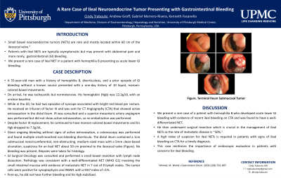
Case Description/Methods: A 55-year-old man with a history of hemophilia B, diverticulosis, and a prior episode of GI bleeding without a known source presented with a one-day history of 10 liquid, maroon-colored bowel movements. On arrival, he was tachycardic but normotensive. His hemoglobin (Hgb) was 12.2g/dL with an unclear baseline. While in the ED, he had two episodes of syncope associated with bright red blood per rectum. He received an infusion of factor IX and was sent for CT Angiography (CTA). There was evidence of blood in the distal ileum without active extravasation and no embolization was performed. Despite factor IX replacement, he continued to have maroon-colored bowel movements and his Hgb dropped to 7.5g/dL. Given ongoing bleeding without signs of active extravasation, a colonoscopy was performed and found multiple small-mouthed non-bleeding diverticula. The distal ileum contained a 1cm submucosal noncircumferential non-obstructing medium-sized mass with a 5mm clean-based ulceration, suspicious for an ileal NET about 50cm proximal to the ileocecal valve (Figure). No bleeding was present. Biopsies were taken for histology. GI Surgical Oncology was consulted and performed a small bowel resection with lymph node dissection. Pathology was consistent with a well-differentiated NET (WHO G1) involving the small intestinal mucosa with evidence of metastatic NET in 7 out of 8 lymph nodes. The tumor cells were positive for synaptophysin and INSM1 with a KI67 index of < 1%. Post-op, he did not have further bleeding and his Hgb stabilized.
Discussion: We present a rare case of a patient with hemophilia B who developed acute lower GI bleeding with evidence of recent ileal bleeding on CTA and was found to have a well-differentiated NET. He then underwent surgical resection which is crucial in the management of ileal NETs as the rate of metastatic disease is ~50%.1 A high index of suspicion for ileal NETs is required in patients with signs of ileal bleeding on CTA for a timely diagnosis. This case reinforces the importance of endoscopic evaluation in patients with concerns for ileal bleeding.
1Ahmed, M. World J Gastrointest Oncol. 2020;12(8):791-807.

Figure: Endoscopic Images of Terminal Ileum Submucosal Tumor
Disclosures:
Cindy Traboulsi indicated no relevant financial relationships.
Andrew Groff indicated no relevant financial relationships.
Gabriel Marrero-Rivera indicated no relevant financial relationships.
Kenneth Fasanella indicated no relevant financial relationships.
Cindy Traboulsi, MD, Andrew Groff, MD, Gabriel Marrero-Rivera, MD, Kenneth Fasanella, MD. P4222 - A Rare Case of Ileal Neuroendocrine Tumor Presenting With Gastrointestinal Bleeding, ACG 2024 Annual Scientific Meeting Abstracts. Philadelphia, PA: American College of Gastroenterology.

