Tuesday Poster Session
Category: GI Bleeding
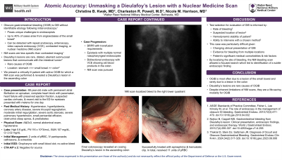
P4224 - Atomic Accuracy: Unmasking a Dieulafoy’s Lesion with a Nuclear Medicine Scan
Tuesday, October 29, 2024
10:30 AM - 4:00 PM ET
Location: Exhibit Hall E

Has Audio
- CK
Christine B. Kwak, MD
Walter Reed National Military Medical Center
Rockville, MD
Presenting Author(s)
Christine B. Kwak, MD1, Charleston R. Powell, MD2, Nicole M. Harrison, MD2
1Walter Reed National Military Medical Center, Rockville, MD; 2Walter Reed National Military Medical Center, Bethesda, MD
Introduction: Obscure gastrointestinal bleeding (OGIB), GIB without identifiable etiology following initial endoscopy, poses unique challenges to endoscopists. OGIB can be detected with repeat endoscopy, enteroscopy, video capsule endoscopy (VCE), contrasted imaging or nuclear medicine (NM) bleeding scans. Dieulafoy’s lesions are rare causes for OGIB usually found in the stomach, occasionally in the small bowel and sparsely in the colon. NM bleeding scans are less specific than contrasted imaging. We present a critically ill patient with active OGIB for which a NM bleeding scan was performed and revealed a Dieulafoy’s lesion in the ascending colon.
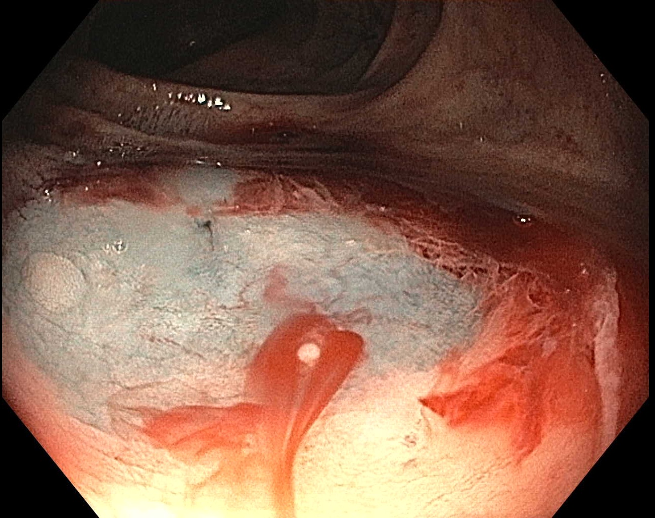
Case Description/Methods: An 88-year-old male with atrial fibrillation on apixaban, congestive heart failure with suspected cardiac cirrhosis, and recent visit to the ED for epistaxis presented with melena for one day. He was hypotensive with MAPs in the 50s. Labs were notable for Hgb of 5.6 g/dL, Plt 116 x 103/mcL, BUN 107 mg/dL, and Cr 2.02 mg/dL. He was transfused, given IV PPI, IV antibiotics, and octreotide. Initial EGD was notable for a small blood clot in the oropharynx. He developed bright red blood per rectum with continued transfusion requirements. He had epistaxis and underwent nasopharyngeal endoscopy that was normal. Bidirectional endoscopy with VCE showed old blood products in the colon. He had ongoing bleeding, not seen on CTA. NM bleeding scan localized bleed to the right lower quadrant and the patient ultimately was found to have an oozing Dieulafoy’s lesion in the ascending colon that was successfully treated with epinephrine and hemostatic clip. In total, he received 11 units of PRBCs.
Discussion: Test selection for evaluation of GIB is informed by the rate of bleeding, suspected location of lesion, hemodynamic stability of patient, and ability to intervene with a chosen method. In this case, the patient had clinical findings and risk factors that made it difficult to focus the evaluation and increased the risk of each possible intervention. By localizing the site of bleeding to the right colon, the NM bleeding scan allowed a focused search which led to identification of a subtle endoscopic finding. Despite inherent limitations of NM scans, it proves to be a life-saving modality for OGIB.

Disclosures:
Christine B. Kwak, MD1, Charleston R. Powell, MD2, Nicole M. Harrison, MD2. P4224 - Atomic Accuracy: Unmasking a Dieulafoy’s Lesion with a Nuclear Medicine Scan, ACG 2024 Annual Scientific Meeting Abstracts. Philadelphia, PA: American College of Gastroenterology.
1Walter Reed National Military Medical Center, Rockville, MD; 2Walter Reed National Military Medical Center, Bethesda, MD
Introduction: Obscure gastrointestinal bleeding (OGIB), GIB without identifiable etiology following initial endoscopy, poses unique challenges to endoscopists. OGIB can be detected with repeat endoscopy, enteroscopy, video capsule endoscopy (VCE), contrasted imaging or nuclear medicine (NM) bleeding scans. Dieulafoy’s lesions are rare causes for OGIB usually found in the stomach, occasionally in the small bowel and sparsely in the colon. NM bleeding scans are less specific than contrasted imaging. We present a critically ill patient with active OGIB for which a NM bleeding scan was performed and revealed a Dieulafoy’s lesion in the ascending colon.
Case Description/Methods: An 88-year-old male with atrial fibrillation on apixaban, congestive heart failure with suspected cardiac cirrhosis, and recent visit to the ED for epistaxis presented with melena for one day. He was hypotensive with MAPs in the 50s. Labs were notable for Hgb of 5.6 g/dL, Plt 116 x 103/mcL, BUN 107 mg/dL, and Cr 2.02 mg/dL. He was transfused, given IV PPI, IV antibiotics, and octreotide. Initial EGD was notable for a small blood clot in the oropharynx. He developed bright red blood per rectum with continued transfusion requirements. He had epistaxis and underwent nasopharyngeal endoscopy that was normal. Bidirectional endoscopy with VCE showed old blood products in the colon. He had ongoing bleeding, not seen on CTA. NM bleeding scan localized bleed to the right lower quadrant and the patient ultimately was found to have an oozing Dieulafoy’s lesion in the ascending colon that was successfully treated with epinephrine and hemostatic clip. In total, he received 11 units of PRBCs.
Discussion: Test selection for evaluation of GIB is informed by the rate of bleeding, suspected location of lesion, hemodynamic stability of patient, and ability to intervene with a chosen method. In this case, the patient had clinical findings and risk factors that made it difficult to focus the evaluation and increased the risk of each possible intervention. By localizing the site of bleeding to the right colon, the NM bleeding scan allowed a focused search which led to identification of a subtle endoscopic finding. Despite inherent limitations of NM scans, it proves to be a life-saving modality for OGIB.

Figure: Oozing Dieulafoy’s lesion in the ascending colon
Disclosures:
Christine Kwak indicated no relevant financial relationships.
Charleston Powell indicated no relevant financial relationships.
Nicole Harrison indicated no relevant financial relationships.
Christine B. Kwak, MD1, Charleston R. Powell, MD2, Nicole M. Harrison, MD2. P4224 - Atomic Accuracy: Unmasking a Dieulafoy’s Lesion with a Nuclear Medicine Scan, ACG 2024 Annual Scientific Meeting Abstracts. Philadelphia, PA: American College of Gastroenterology.

