Tuesday Poster Session
Category: General Endoscopy
P4132 - The Gut Feeling: Unraveling the Quest for the Origin of Tumor Marker
Tuesday, October 29, 2024
10:30 AM - 4:00 PM ET
Location: Exhibit Hall E

Has Audio

Maha Zafar, MD
Mercy Hospital Fort Smith
Fort Smith, AR
Presenting Author(s)
Maha Zafar, MD1, Aswanth Reddy, MD2
1Mercy Hospital Fort Smith, Fort Smith, AR; 2Mercy Hospital, Fort Smith, AR
Introduction: Breast cancer is the leading cause of cancer-related deaths for women aged 20-59. Invasive lobular cancer, accounts for 10 % of all breast cancers. These cancers can manifest at atypical metastatic sites including bone marrow and peritoneum, distinguishing them from ductal cancers. This case report represents a rare instance of gastrointestinal metastasis featuring elevated tumor markers in a previously asymptomatic patient.
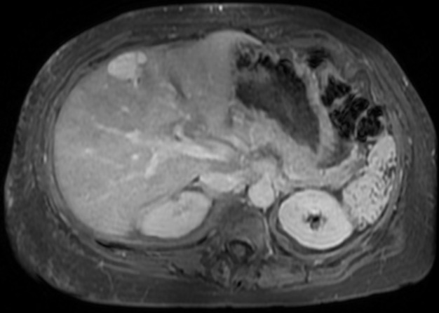
Case Description/Methods: A 52-year-old woman with a history of left-sided infiltrating lobular breast cancer was referred to our oncology clinic for elevated tumor markers. Previously, she had bilateral mastectomy and axillary lymph node dissection with final stage of IIIC (T1cN3M0). She declined adjuvant chemotherapy, radiation therapy and started anastrozole 1mg daily. Surveillance laboratory tests showed elevated liver function tests and tumor markers CA-125 and CA 15-3. PET scan showed no malignancy. Ultrasound liver revealed fatty liver with irregular contours, and lobulated cyst on liver MRI. Three months later, she was evaluated for new-onset diarrhea. Upper GI endoscopy and colonoscopy showed mildly edematous mucosa in duodenum and colon without any obvious lesions. Multiple random biopsies revealed metastatic breast cancer in duodenum, transverse, and descending colon, with strong positivity to estrogen and progesterone receptors and negative for HER2. She was then started on systemic therapy with a cyclin-dependent kinase (CDK) 4/6 inhibitor in addition to anastrozole.
Discussion: Breast cancer is most common cancer affecting females globally, with 2.4 million new cases annually. GI metastasis is rare but is primarily linked to invasive lobular carcinoma. The stomach typically emerges as primary site, with involvement of small or large intestine being rare. Our case introduced a compelling twist with metastasis of both the small and large intestine. Notably, endoscopic findings unveiled an edematous mucosa, confirming submucosal involvement and underscoring the significance of random biopsies. The challenge lies in non-specific nature of GI metastasis symptoms, potentially leading to delayed diagnosis and treatment, thus adversely impacting prognosis. Despite its rarity, GI metastasis should remain on the radar for patients with a history of breast cancer presenting with ambiguous GI symptoms or elevated tumor markers, as observed in our case. This proactive approach promotes prompt detection and management of potential GI metastasis, improving patient outcomes.

Disclosures:
Maha Zafar, MD1, Aswanth Reddy, MD2. P4132 - The Gut Feeling: Unraveling the Quest for the Origin of Tumor Marker, ACG 2024 Annual Scientific Meeting Abstracts. Philadelphia, PA: American College of Gastroenterology.
1Mercy Hospital Fort Smith, Fort Smith, AR; 2Mercy Hospital, Fort Smith, AR
Introduction: Breast cancer is the leading cause of cancer-related deaths for women aged 20-59. Invasive lobular cancer, accounts for 10 % of all breast cancers. These cancers can manifest at atypical metastatic sites including bone marrow and peritoneum, distinguishing them from ductal cancers. This case report represents a rare instance of gastrointestinal metastasis featuring elevated tumor markers in a previously asymptomatic patient.
Case Description/Methods: A 52-year-old woman with a history of left-sided infiltrating lobular breast cancer was referred to our oncology clinic for elevated tumor markers. Previously, she had bilateral mastectomy and axillary lymph node dissection with final stage of IIIC (T1cN3M0). She declined adjuvant chemotherapy, radiation therapy and started anastrozole 1mg daily. Surveillance laboratory tests showed elevated liver function tests and tumor markers CA-125 and CA 15-3. PET scan showed no malignancy. Ultrasound liver revealed fatty liver with irregular contours, and lobulated cyst on liver MRI. Three months later, she was evaluated for new-onset diarrhea. Upper GI endoscopy and colonoscopy showed mildly edematous mucosa in duodenum and colon without any obvious lesions. Multiple random biopsies revealed metastatic breast cancer in duodenum, transverse, and descending colon, with strong positivity to estrogen and progesterone receptors and negative for HER2. She was then started on systemic therapy with a cyclin-dependent kinase (CDK) 4/6 inhibitor in addition to anastrozole.
Discussion: Breast cancer is most common cancer affecting females globally, with 2.4 million new cases annually. GI metastasis is rare but is primarily linked to invasive lobular carcinoma. The stomach typically emerges as primary site, with involvement of small or large intestine being rare. Our case introduced a compelling twist with metastasis of both the small and large intestine. Notably, endoscopic findings unveiled an edematous mucosa, confirming submucosal involvement and underscoring the significance of random biopsies. The challenge lies in non-specific nature of GI metastasis symptoms, potentially leading to delayed diagnosis and treatment, thus adversely impacting prognosis. Despite its rarity, GI metastasis should remain on the radar for patients with a history of breast cancer presenting with ambiguous GI symptoms or elevated tumor markers, as observed in our case. This proactive approach promotes prompt detection and management of potential GI metastasis, improving patient outcomes.

Figure: Figure 1, MRI abdomen W WO contrast revealing a 3 x 2.5 cm lobulated cyst at the junction of right and left liver lobes.
Disclosures:
Maha Zafar indicated no relevant financial relationships.
Aswanth Reddy indicated no relevant financial relationships.
Maha Zafar, MD1, Aswanth Reddy, MD2. P4132 - The Gut Feeling: Unraveling the Quest for the Origin of Tumor Marker, ACG 2024 Annual Scientific Meeting Abstracts. Philadelphia, PA: American College of Gastroenterology.
