Tuesday Poster Session
Category: General Endoscopy
P4134 - Appendiceal Adenocarcinoma Diagnosed via Colonoscopy and Biopsy of the Appendiceal Orifice
Tuesday, October 29, 2024
10:30 AM - 4:00 PM ET
Location: Exhibit Hall E

Has Audio

Akshay Duddu, MD
Greater Baltimore Medical Center
Towson, MD
Presenting Author(s)
Akshay Duddu, MD1, Jayashrei Sairam, MD1, M Kenan Rahima, MD2, Mohammed Rifat Shaik, MBBS3, Rahul Sharma, MD1, Niraj Jani, MD1
1Greater Baltimore Medical Center, Towson, MD; 2TriHealth Good Samaritan Hospital, Cincinnati, OH; 3University of Maryland Medical Center Midtown Campus, Baltimore, MD
Introduction: Appendiceal cancer is very rare. In the United States (US), it has an estimated incidence of 1 - 2 cases/million people/year. In recent years, however, the incidence is on the rise. Mucinous adenocarcinoma of the appendix is an epithelial variant most often diagnosed post-operatively in appendectomy specimens of patients who were initially diagnosed with appendicitis. We present an exceedingly rare case where endoscopic visualization of the appendiceal orifice hinted at the diagnosis of an appendiceal adenocarcinoma.
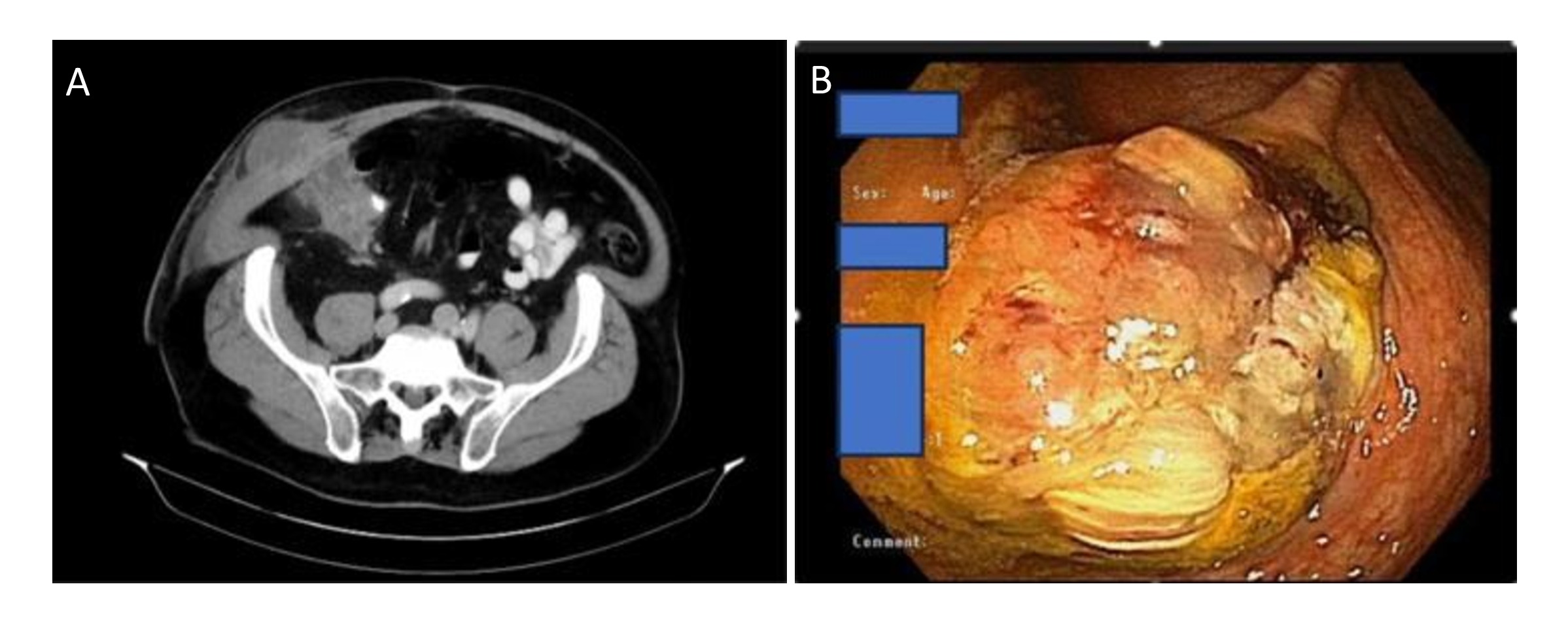
Case Description/Methods: A 78-year-old gentleman with a history of appendiceal rupture complicated by phlegmon formation managed with laparoscopic washout and drainage, JP drain placement, and antibiotics 8 months ago presented to the hospital with worsening right lower quadrant (RLQ) abdominal pain and swelling that started a week ago. An 8 x 6 cm tender mass was palpable in the RLQ with a leukocytosis of 17,000. A CT scan of the abdomen and pelvis with contrast revealed a large heterogeneous mass in the RLQ measuring 10 x 5 cm extending to the abdominal wall, suggesting either an inflammatory process or malignancy. The patient was initiated on IV antibiotics and gastroenterology was consulted for endoscopic evaluation of the terminal ileum and cecum given concern for a neoplastic process. Colonoscopy revealed ulcerated, non-bleeding, nodular appearance of the mucosa at the appendiceal orifice with significant mucin extrusion. Biopsy revealed in-situ and invasive moderately differentiated adenocarcinoma. Surgical oncology was consulted, and en-bloc resection of the anterior abdominal wall mass with ileo-colectomy was performed. Surgical pathology of the resected mass again confirmed the diagnosis.
Discussion: Appendiceal adenocarcinoma is an extremely rare diagnosis in the general population. However, the prevalence of appendiceal cancers in appendectomy specimens for patients presenting with acute appendicitis can be as high as 1.9%. The diagnosis of appendiceal cancer can be missed or delayed if an appendectomy is not performed due to local complications such as phlegmon formation, as in our case. Fortunately, the endoscopic evaluation of the appendiceal orifice and its biopsy led to an early discovery and curative treatment. Our case highlights the diagnostic value of colonoscopy and appendiceal orifice examination in the early identification of appendiceal cancers in patients who could not undergo initial appendectomy.

Disclosures:
Akshay Duddu, MD1, Jayashrei Sairam, MD1, M Kenan Rahima, MD2, Mohammed Rifat Shaik, MBBS3, Rahul Sharma, MD1, Niraj Jani, MD1. P4134 - Appendiceal Adenocarcinoma Diagnosed via Colonoscopy and Biopsy of the Appendiceal Orifice, ACG 2024 Annual Scientific Meeting Abstracts. Philadelphia, PA: American College of Gastroenterology.
1Greater Baltimore Medical Center, Towson, MD; 2TriHealth Good Samaritan Hospital, Cincinnati, OH; 3University of Maryland Medical Center Midtown Campus, Baltimore, MD
Introduction: Appendiceal cancer is very rare. In the United States (US), it has an estimated incidence of 1 - 2 cases/million people/year. In recent years, however, the incidence is on the rise. Mucinous adenocarcinoma of the appendix is an epithelial variant most often diagnosed post-operatively in appendectomy specimens of patients who were initially diagnosed with appendicitis. We present an exceedingly rare case where endoscopic visualization of the appendiceal orifice hinted at the diagnosis of an appendiceal adenocarcinoma.
Case Description/Methods: A 78-year-old gentleman with a history of appendiceal rupture complicated by phlegmon formation managed with laparoscopic washout and drainage, JP drain placement, and antibiotics 8 months ago presented to the hospital with worsening right lower quadrant (RLQ) abdominal pain and swelling that started a week ago. An 8 x 6 cm tender mass was palpable in the RLQ with a leukocytosis of 17,000. A CT scan of the abdomen and pelvis with contrast revealed a large heterogeneous mass in the RLQ measuring 10 x 5 cm extending to the abdominal wall, suggesting either an inflammatory process or malignancy. The patient was initiated on IV antibiotics and gastroenterology was consulted for endoscopic evaluation of the terminal ileum and cecum given concern for a neoplastic process. Colonoscopy revealed ulcerated, non-bleeding, nodular appearance of the mucosa at the appendiceal orifice with significant mucin extrusion. Biopsy revealed in-situ and invasive moderately differentiated adenocarcinoma. Surgical oncology was consulted, and en-bloc resection of the anterior abdominal wall mass with ileo-colectomy was performed. Surgical pathology of the resected mass again confirmed the diagnosis.
Discussion: Appendiceal adenocarcinoma is an extremely rare diagnosis in the general population. However, the prevalence of appendiceal cancers in appendectomy specimens for patients presenting with acute appendicitis can be as high as 1.9%. The diagnosis of appendiceal cancer can be missed or delayed if an appendectomy is not performed due to local complications such as phlegmon formation, as in our case. Fortunately, the endoscopic evaluation of the appendiceal orifice and its biopsy led to an early discovery and curative treatment. Our case highlights the diagnostic value of colonoscopy and appendiceal orifice examination in the early identification of appendiceal cancers in patients who could not undergo initial appendectomy.

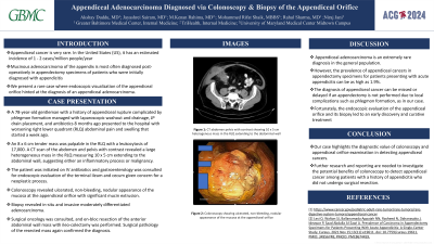
Figure: Figure A: CT abdomen pelvis with contrast showing 10 x 5 cm heterogenous mass in the RLQ extending to the abdominal wall
Figure B: Colonoscopy showing ulcerated, non-bleeding, nodular appearance of the mucosa at the appendiceal orifice
Figure B: Colonoscopy showing ulcerated, non-bleeding, nodular appearance of the mucosa at the appendiceal orifice
Disclosures:
Akshay Duddu indicated no relevant financial relationships.
Jayashrei Sairam indicated no relevant financial relationships.
M Kenan Rahima indicated no relevant financial relationships.
Mohammed Rifat Shaik indicated no relevant financial relationships.
Rahul Sharma indicated no relevant financial relationships.
Niraj Jani indicated no relevant financial relationships.
Akshay Duddu, MD1, Jayashrei Sairam, MD1, M Kenan Rahima, MD2, Mohammed Rifat Shaik, MBBS3, Rahul Sharma, MD1, Niraj Jani, MD1. P4134 - Appendiceal Adenocarcinoma Diagnosed via Colonoscopy and Biopsy of the Appendiceal Orifice, ACG 2024 Annual Scientific Meeting Abstracts. Philadelphia, PA: American College of Gastroenterology.
