Tuesday Poster Session
Category: General Endoscopy
P4135 - Sapphire Shadows: A Case Series Describing Unusual Ileal Discoloration in a Veteran Population
Tuesday, October 29, 2024
10:30 AM - 4:00 PM ET
Location: Exhibit Hall E

Has Audio
- LT
Lyubov Tiegs, MD
University of Minnesota
Minneapolis, MN
Presenting Author(s)
Lyubov Tiegs, MD1, Bryant Megna, MD2, Justin Peltola, MD3, Daniela Guerrero Vinsard, MD3
1University of Minnesota, Minneapolis, MN; 2University of Minnesota Medical Center, Minneapolis, MN; 3Minneapolis VA Health Care System, Minneapolis, MN
Introduction: Existing literature has described the presence of black granular pigment deposits in the base of Peyer patches of the terminal ileum, presumed to be secondary to macrophage uptake of microparticles. However, large spots of pigmentation grossly visible on routine colonoscopy with ileal intubation have not been reported. We present four cases of visible blue pigmentation in the ileum of curious etiology.
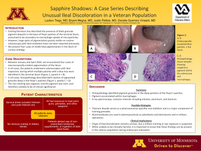
Case Description/Methods: Between January and April 2024, we encountered four cases of macroscopically evident pigmentation of the ileum. In all cases, the patients underwent colonoscopies with ileal inspection during which multiple patches with a blue hue were identified in the terminal ileum (Figure 1, A + B). In all cases, histopathology described dark clusters of pigmented granules deep in the Peyer’s patches (Figure 2, C + D). The iron staining was negative, and the pigment was inert, and therefore unlikely to be of clinical significance. The patients were subsequently interviewed, and their records examined revealing the following commonalities. All patients were veterans, their service times including the Vietnam and post-Vietnam era. Although they served in different areas, on interview they endorsed exposure to lead paint during their service time, as well as exposure to guns, grenades and other weapons during basic training. There was no obvious overlap of dietary trends. The patients denied the use of non-prescribed medicines or supplements and denied ingestion of dark-dyed foods.
Discussion: In all four cases, histopathology identified pigment granules in the deep portions of the Peyer’s patches. Pathology reports described that the pigment accumulated within macrophages and has been shown by x-ray spectroscopy to contain mineral composition that includes silicates, aluminum, and titanium, with the most plausible explanation being of atmospheric or dietary source. Literature review revealed that titanium dioxide serves as an opacifier and stabilizer in pharmaceuticals and is a major component of training grenades. Aluminosilicates are used in the pharmaceuticals as adsorbents and demulcents and in military operations. Ultimately, the primary cause of blue patches in the ileum remains unclear, but given the military background of the patients, a training or war-related exposure could be presumed. While unlikely clinically harmful, it is important to know that these findings can be present in the veteran population during endoscopic evaluation.

Disclosures:
Lyubov Tiegs, MD1, Bryant Megna, MD2, Justin Peltola, MD3, Daniela Guerrero Vinsard, MD3. P4135 - Sapphire Shadows: A Case Series Describing Unusual Ileal Discoloration in a Veteran Population, ACG 2024 Annual Scientific Meeting Abstracts. Philadelphia, PA: American College of Gastroenterology.
1University of Minnesota, Minneapolis, MN; 2University of Minnesota Medical Center, Minneapolis, MN; 3Minneapolis VA Health Care System, Minneapolis, MN
Introduction: Existing literature has described the presence of black granular pigment deposits in the base of Peyer patches of the terminal ileum, presumed to be secondary to macrophage uptake of microparticles. However, large spots of pigmentation grossly visible on routine colonoscopy with ileal intubation have not been reported. We present four cases of visible blue pigmentation in the ileum of curious etiology.
Case Description/Methods: Between January and April 2024, we encountered four cases of macroscopically evident pigmentation of the ileum. In all cases, the patients underwent colonoscopies with ileal inspection during which multiple patches with a blue hue were identified in the terminal ileum (Figure 1, A + B). In all cases, histopathology described dark clusters of pigmented granules deep in the Peyer’s patches (Figure 2, C + D). The iron staining was negative, and the pigment was inert, and therefore unlikely to be of clinical significance. The patients were subsequently interviewed, and their records examined revealing the following commonalities. All patients were veterans, their service times including the Vietnam and post-Vietnam era. Although they served in different areas, on interview they endorsed exposure to lead paint during their service time, as well as exposure to guns, grenades and other weapons during basic training. There was no obvious overlap of dietary trends. The patients denied the use of non-prescribed medicines or supplements and denied ingestion of dark-dyed foods.
Discussion: In all four cases, histopathology identified pigment granules in the deep portions of the Peyer’s patches. Pathology reports described that the pigment accumulated within macrophages and has been shown by x-ray spectroscopy to contain mineral composition that includes silicates, aluminum, and titanium, with the most plausible explanation being of atmospheric or dietary source. Literature review revealed that titanium dioxide serves as an opacifier and stabilizer in pharmaceuticals and is a major component of training grenades. Aluminosilicates are used in the pharmaceuticals as adsorbents and demulcents and in military operations. Ultimately, the primary cause of blue patches in the ileum remains unclear, but given the military background of the patients, a training or war-related exposure could be presumed. While unlikely clinically harmful, it is important to know that these findings can be present in the veteran population during endoscopic evaluation.

Figure: Figure 1
A, B: Endoscopically visualized blue patches in the ileum.
C, D: Histopathology tissue samples showing exogenous pigment within the submucosa and macrophages.
A, B: Endoscopically visualized blue patches in the ileum.
C, D: Histopathology tissue samples showing exogenous pigment within the submucosa and macrophages.
Disclosures:
Lyubov Tiegs indicated no relevant financial relationships.
Bryant Megna indicated no relevant financial relationships.
Justin Peltola indicated no relevant financial relationships.
Daniela Guerrero Vinsard indicated no relevant financial relationships.
Lyubov Tiegs, MD1, Bryant Megna, MD2, Justin Peltola, MD3, Daniela Guerrero Vinsard, MD3. P4135 - Sapphire Shadows: A Case Series Describing Unusual Ileal Discoloration in a Veteran Population, ACG 2024 Annual Scientific Meeting Abstracts. Philadelphia, PA: American College of Gastroenterology.
